qualified by the words ‘if necessary’. Whereas some fractures must be splinted
rigidly, many do not require immobilisation to ensure union, and excessive
immobilisation is actually harmful in some (Figs 3.2 & 3.3).
INDICATIONS FOR IMMOBILISATION
There are only three reasons for immobilising a fracture:
1. to prevent displacement or angulation of the fragments
2. to prevent movement that might interfere with union
3. to relieve pain.
Prevention of displacement or angulation
As a general rule, the broken fragments will not become displaced more
severely than they were at the time of the original injury. Therefore, if the
original position is acceptable, immobilisation to prevent further displacement
is often unnecessary. In fractures of the shafts of the major long bones, however,
immobilisation is usually necessary in order to maintain correct alignment.
Prevention of movement
As has been mentioned already, absolute immobility is not always essential to
union of a fracture. It is only when movement might shear the delicate capillaries bridging the fracture that it is undesirable, and, theoretically, rotation
movements are worst in this respect. There are three fractures that constantly
demand immobilisation to ensure their union—namely, those of the scaphoid
bone, of the shaft of the ulna, and of the neck of the femur.
Examples of fractures that heal well without immobilisation are those of
the ribs, clavicle and scapula, and stable fractures of the pelvic ring.
Immobilisation is also unnecessary for certain fractures of the humerus and
femur, and many fractures of the metacarpals, metatarsals and phalanges. In
some fractures, excessive immobilisation may do more harm than good. The
injured hand, in particular, tolerates prolonged immobilisation badly. Whereas
the wrist may be immobilised for many weeks or even months with impunity,
to immobilise injured fingers for a long time is to court disaster in the form of
permanent joint stiffness.
Relief of pain
Probably in about half of all the cases in which a fracture is immobilised the
main reason for immobilisation is to relieve pain. With the limb thus made
comfortable, it can be used much more effectively than would otherwise be
possible.
METHODS OF IMMOBILISATION
When immobilisation is deemed necessary there are four methods by which it
may be effected:
1. by a plaster of Paris cast or other external splint
2. by continuous traction
3. by external fixation
4. by internal fixation.
Immobilisation by plaster, splint or brace
For most fractures the standard method of immobilisation is by a plaster of
Paris cast. Also available are various proprietary substitutes for plaster, which
offer the advantages of lighter weight, radiolucency and imperviousness to
water, though at much greater cost. Most such products are also more difficult
to apply; nevertheless, they are being used on an increasing scale. For some
fractures a splint made from metal, wood or plastic is more appropriate—for
example, the Thomas’s splint for fractures of the shaft of the femur, or a plastic
collar for certain injuries of the cervical spine.
Plaster technique. Plaster of Paris is hemihydrated calcium sulphate. It reacts
with water to form hydrated calcium sulphate. The reaction is exothermic, a
fact that is evidenced by noticeable warming of the plaster during setting.
Plaster bandages may be prepared by impregnating rolls of book muslin
with the dry powdered plaster, but except in a few developing countries, most
hospitals now use ready-made proprietary bandages. These are best used with
cold water because setting is too rapid with warm water.
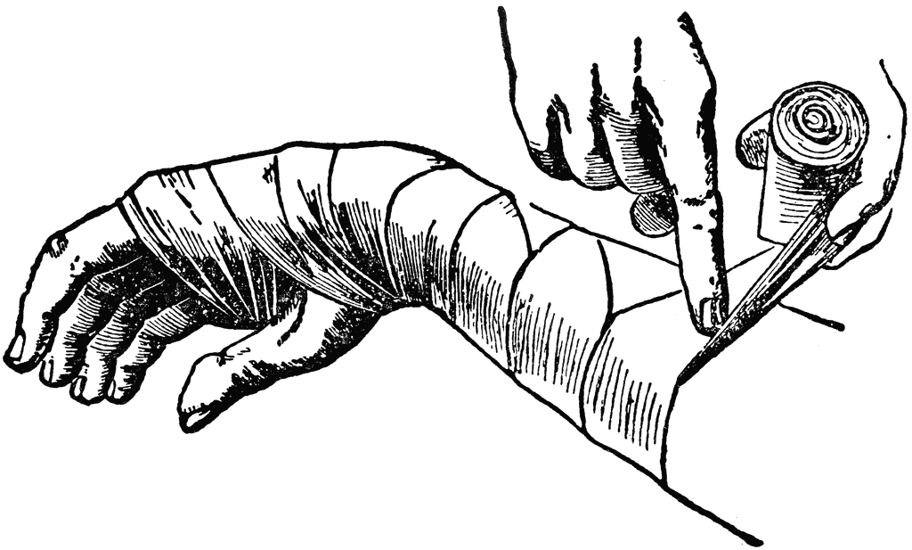
Most surgeons use a thin lining of stockinet or cellulose bandage to prevent
the plaster from sticking to the hairs and skin (Fig. 3.4). The use of a lining is
certainly recommended because it adds greatly to the comfort of the plaster. If
marked swelling is expected, as after an operation upon the limb, a more bulky
padding of surgical cotton wool should be used.
The plaster bandages are applied in two forms: round-and-round bandages
and longitudinal strips or ‘slabs’ to reinforce a particular area. Round-andround bandages must be applied smoothly without tension, the material being
drawn out to its full width at each turn. Slabs are prepared by unrolling a
bandage to and fro upon a table: an average slab consists of about 12 thicknesses. The slabs are placed at points of weakness or stress and are held in
place by further turns of plaster bandage.
A plaster is best dried simply by exposure to the air: artificial heating is
unnecessary. A plaster will not dry satisfactorily if it is kept covered by clothing
or bed-linen.
Synthetic (plastic) splinting materials are applied in much the same way as
plaster bandages, usually with warm water. Since they are stronger weight for
weight than plaster, fewer layers are required. Moulding to the body contours
is more difficult than with plaster bandages.
Immobilisation by internal fixation
Operative or internal fixation may be advised in the following circumstances:
1. to provide early control of limb fractures when conservative methods
would interfere with the management of other severe injuries, for instance
of the head, thorax or abdomen
2. as a method of choice in certain fractures, to secure immobilisation of the
fracture and to allow early mobility of the patient, e.g. in the elderly
patient with trochanteric hip fracture
3. when it has been necessary to operate upon a fracture to secure adequate
reduction
4. if it is impossible in a closed fracture to maintain an acceptable position by
splintage alone.
Methods of internal fixation. The following methods are currently in general
use (Fig. 3.16):
1. metal plate held by screws or locking plate (with screws fixed to the plate
by threaded holes)
2. intramedullary nail, with or without cross-screw fixation for locking
3. dynamic compression screw-plate
4. condylar screw-plate
5. tension band wiring
6. transfixion screws.
The choice of method depends upon the site and pattern of the fracture.
Plate and screws. This method is applicable to long bones. Usually a single
six-hole plate suffices, but an eight-hole plate may be preferred for larger bones.
Fixation by ordinary plates has the disadvantage that the bone fragments
are not forcibly pressed into close contact; indeed, if there is any absorption of the fracture surfaces the plate tends to hold the fragments apart, and this may
sometimes be a factor in the causation of delayed union. In order to counter this
disadvantage of simple plates and to improve coaptation at the time of plating,
special compression plates are available by which the fragments are forced
together before the plate is finally screwed home (compression plating).
Locking plate. A newer concept is the ‘locking plate’, that uses screws with
heads that are threaded and when tightened lock into matching threads in the
holes of the plate. This produces a more rigid fixation in terms of length and
angle, which is particularly valuable in comminuted fractures in osteoporotic
bone. It can also be inserted with less stripping of soft tissue that preserves
bone vascularity, particularly in the metaphyseal region.
Intramedullary nail. This technique is excellent for many fractures of the long
bones, especially when the fracture is near the middle of the shaft. It is used
regularly for fractures of the femur and tibia, and less commonly in the
humerus. The original Kuntscher-type nail designed for the femur was hollow
and of clover-leaf section and achieved fixation by its tight fit in the narrowest
isthmus of the shaft. This type has been replaced by the newer more versatile
locking nail with a rounder cross-section (Fig. 3.16), which offers notable
advantages. These have transverse holes at both ends, allowing the insertion of
transfixion (‘locking’) screws through bone and nail under image intensifier
radiographic control. This affords greater rigidity as well as resistance to
rotation forces allowing their use in comminuted fractures, particularly in the
wider medullary canal near the bone ends. A new design of thinner more flexible
solid nail is sometimes used for the management of shaft fractures in children.
Compression screw-plate. The compression screw-plate (dynamic hip screw) is
a standard method of fixation for fractures of the neck of the femur and for
trochanteric fractures (see Fig. 15.3). The screw component, which grips the
femoral head, slides telescopically in the barrel to allow the bone fragments to
be compressed together across the fracture. This compression effect is brought
about by tightening a screw in the base of the barrel.
Transfixion screws. The use of a transfixion screw has wide application in the
fixation of small detached fragments—for instance the capitulum of the
humerus, the olecranon process of the ulna or the medial malleolus of the tibia.
Kirschner wire fixation. These thin flexible wires with sharpened ends are
available in a number of diameters and provide a useful alternative to
transfixion screws for the fixation of small bony fragments or for fractures of
the small bones in the hand and foot.
Tension band wiring. This technique of fixation is most commonly used in the
patella and olecranon, but can be applied to other small metaphyseal fragments
such as the medial malleolus. It uses the mechanical principle of converting the
tensile stresses of the muscles acting on the bone fragment, into a compressive
force at the fracture site. This is achieved by means of tightening an eccentric
figure-of-eight cerclage wire across the two fragments, stabilised by Kirschner
wires or a screw inserted at right angles to the fracture line (Fig. 3.16).
Metals for internal fixation
Metals used for internal fixation of fractures or for internal prostheses must be
resistant to corrosion in the tissues: silver, iron, ordinary steel and nickel-plated
steel are all unsuitable. A special stainless steel containing chromium, nickel
and molybdenum is widely used, but a non-ferrous alloy containing
chromium, cobalt and molybdenum has even better resistance to corrosion in
the body and is used for all types of internal appliance except wire, for whichit is technically unsuitable. The metallic element titanium and its alloys have
also proved resistant to corrosion in the body and are used increasingly for the
manufacture of prostheses and internal fixation devices.
The place of operative fixation
In recent years there has been an increasing use of internal fixation for the
treatment of limb fractures in most trauma centres, often as a deliberate first
choice. As will be seen in a later chapter, operative fixation is already accepted
as the best routine method of treating fractures of the neck and trochanteric
region of the femur in the elderly. Until recently, many fractures of the shafts of
the long bones have been treated conservatively—generally with excellent
results, although often at the cost of rather a long time in hospital or away from
work. The introduction of more sophisticated implants, inserted through small
incisions under radiological screening, and offering immediate fracture fixation,
has led to a dramatic change in this policy. In particular, intramedullary nailing
is now used for most fractures of the shaft of the femur or tibia.
The reasons for advocating surgical intervention for fractures that were
formerly managed conservatively are threefold. Firstly, there may be a
substantial reduction in the time that the patient must spend in hospital and
away from work. Secondly, in a favourable case function of the limb—and
particularly of the joints—may be restored earlier because the need for plaster
or other external splintage can often be eliminated. And thirdly, it is hoped that
by providing rigid fixation of the fracture, complications such as delayed union
and non-union will be reduced. In themselves, these objectives are unexceptionable, but there are arguments on the other side. The chief of these is that
operative fixation, especially when combined with open reduction, entails risks
that are absent or minimal with conservative treatment. Occasional fatalities—
for instance from pulmonary embolism—are probably unavoidable, and major
wound infection is by no means uncommon after lengthy open operations for
reduction and internal fixation. Extensive stripping of soft tissues from the
bone may also lead to adhesions that restrict joint movement, and may jeopardise the blood supply to the bone fragments, thereby hindering union. Thus
the objects of the operation may sometimes be defeated.
It is important to strike a fair balance between these conflicting arguments,
and to weigh up all the factors in every case: the age of the patient, the site and
nature of the fracture, problems of employment, and economic circumstances.
Advanced age should always weigh heavily in favour of an operation that will
enable the patient to get out of bed sooner, whereas anything that might favour
infection, such as an open wound or a pressure blister, should weigh heavily
against open operation. In such cases external fixation (p. 41) as distinct from
internal fixation has an important place.
The final decision on whether to use internal fixation or an alternative
conservative method of fracture treatment must be made by the surgeon. They
must be guided by their experience with the technique, the availability of the
implants, the operating and ward environments, and the incidence of wound
infection in other patients.


No comments:
Post a Comment