ayat gerak

Aku dye..cinta kami..kisah kami..sayang dye..bila kowg dah masuk blog kami..kowg kena gak follow kami tau!!! ^_^

CoR3t@n Kami...,

ASSALAMMUAILAIKUM....
selamat datang kew ruangan kami..,so..,d sbb kn nei ruangan kami..,suka aty kami ler nak tulis entry pew..,n nak letak pew..,klu rasa x suka..,wat xtau jew eak..,i don't care about that!!!!!!! cbb ruangan nei sentiasa nyer ada pic d setiap warkah..,k ler..,hope ur all enjoy k..dah singgah jgn lupa follow n leave ur comment.,,daaa^_^

Thursday 29 September 2011

0
Komen

Pew tuuuuuuuu...???????????

Assalammuailaikum semuaaaa....,,
hye..,pew khabor kowg semua..,hope kowg sihat wafiat ler eak..,kalau sakit tu..,pegi cepat2 jumpa 'abang ensem & akk pretty (doctor lerrr..,)..,utk bik ubat eak.,sekarang nei sye tengah busy ckit ler.,dengan dunia practical ku..,hihihi.,ini pun sye dapat luang kan masa dengan kowg semua sbb shift sye pagi tadi..,soo..,malam nei adalah time bersama kowg semua..,

okey..,sye xmau terlalu panjang mukadimah nyer..,i want to straight the point ler..,about my title okey..,al-kisah nyer cam nei eak kawan2 ku..,di sebabkan hari nei sye shift pagi.,so.,pagi2 buta kena ler membuka mata seluas alam untuk melihat keindahan pagi yang gelap gelita..,seawal 4 pagi bgun semata-mata untuk menjalankan tugasan yang telah di tetapkan..,owg laen sedang enak di ulit mimpi indah,,yang sye lak terpaksa meninggalkan segala keindahan mimpi tu..,tapi kan..,ada gak dalam diam..,seawal pagi tu..,yang smua penghuni sedap2 layan mimpi..,de yang sanggup bertahan mata nyer untuk membuat ''acting'' awal pagi tu..,PEW KE JADAH mrke tu hahhhh!!!!

Ada yang sanggup berjaga semata2 nak buat MAKSIAT..,klu wat maksiat kat luar aq xkesah sngt..,yang aq kesah nyer..,bila owg tu buat oner kat umah yang aq duduki n aq sewa untuk aq menuntut ilmu..,!!! de lak yang gatal dye punye ''anu''..,wat MAKSIAT kat umah nei lak..,!!!! burn jew hati aq..,uuhhh fuuuhhh!!! pandai lak nak usha2 owg lak..,dah buat kesalah xreti rasa malu langsung..,!!! klu aq ler..,dah lama aq sorok muka kat dlm ''MANGKUK JAMBAN'' owg cam tu..,x patut di hormat lagi..,rasa nak terajang2 jew betina jalang tu!!! dye gat dye MAMMM BESAR kat dlm umah neiii..??? !! sesuka hati nenek moyang dye nak bawak jantan masuk umah..,!!!! eeaakkkk!!!!..,panasssss....panassss...,

okey ler kowg..,aq nak tdo dulu..,esok nak bgun awak lagi..,uurrmmm sowi ler eak atas p'ggunaan bahasa yang agak lancang n xteratur tu..,aq dah terbiasa m'bahasa kan diri ngan bahasa aq..,better aq terus kan ngan panggilan tu eak..,hope kowg x kesah..,

MORAL : Kita kalau owg islam..,hormati ler agama kita..,jgn jejas kan image bersih agama kita..,hope owg yg buat benda tu..,ALLAH swt tunjuk kan jln yg benar buat dye..,insyallah..,


Ko Gemar LIKE ler.,xGemar leave COMMENT

Monday 26 September 2011

0
Komen

uuurrmm..,

Assalammuailaikum..,
urrmm..,aq rasa ler..,ary nei aq cam nak edit blog aq jew..,sambil tu nak usha toturial Lyssa faizureen..,nei tau leh d gnukan utk m'hias blog aq yg sntiasa sepi nei..,bkn nyer nak edit pew pown..,juz nak tukar background n yg mne yg x brpew penting tu..,nak di hapuskan lorhh..,uhuhuh..,sambil tu..,nak merewang kat blog2 laen yg de toturial..,tpi xtau sapa lg..,sooo...,yg blog sapa yg bertuah tu eak...????? tgguuuuuuuuuuuuuu........,


Ko Gemar LIKE ler.,xGemar leave COMMENT

Thursday 22 September 2011

0
Komen

Suka Hati Ko Jew..,!!!


Assalammuailaikum semua.., 
Hye..,lama kan aq xmsuk kan entry bru..,klu ada pun..,about knowlegde..,hihihi..,tapi ary nei aq nak share story ler ngan kowg..,sjew wat pedoman..,uurrmm..,kisah nyer mesti x lari..,kalau sakit hati and jiwa jew..,mesti kat bebudak umah ler..,pew g..,urrmm..,aq telah d maklum kan oleh akk blek aq..,bahawa umah yg aq dok nei..,akan digunakan utk jamuan perpisahan n raya utk group mrke..,masalh nyer yg timbul nei..,SUKA2 JEW DEMA TU NAK WAT SUGGESTION..,THEN SKEW2 JEW WAT KEPUTUSAN.,TANPA DISCUSS NGAN BDK UMAH LAEN..,!!!
Xde kne m'gena..,hahahha..,
Uuurrmmmm..,benda ini berlaku di sbbkan oleh BAJET leader kat umah aq tu..,jgn fikir..,''ko leader..,segala hal umah ko leh wat sesuka hati ko jew..,!!!'' otak dah tua..,fikir ler..,nak sound owg pandai.,kata aq x pemikiran matang ler..,jgn terlampau kekampungan ler.,cakap x gne otak ler!!! cam ler ko tu bagus sngt..,uuhhh!!! kalau ko rasa ko tu bagus..,MATANG sngt..,npew nak wat keputusan tu tanpa bincang ngan bdk umah??? n npew sesuka hati ko jew nak cam tu..??? urrmm..,jawapan nyer sng jew..,SELFISH..,tu ler jwpn nyer..,

Aq xkesah ko nak buat pew.,tapi yg aq kesah..,umah tu bukan nyer umah ko sowg..,aq pun bayar sewa..,beratus2 tiap bulan aq bayar.,aq pun de hak kat umah tu..,bg aq ler..,xperlukan leader utk arrange smue tu..,xguna ada leader..,tapi umah cam kapal pecah..,tonggang langgang..,something yg ptt di bwk bncg..,xreti2 nak bawak ketengah kew??? klu hal melibatkan owg dlm umah..,laen crte nyer..,tapi nei akan melibat kan owg luar..,masalahnyer..,ko akan membawa owg luar kew dlm umah!!! ko tu x berotak kew nak fkr.??? klu hal nei antra owg dlm umah jew..,hnye 7 kepala yg akan terlibat..!!! tapi nei akan terlibabt ngan berpuluh2 kepala.!!! di kata kan dlm majlis jamuan tu..,de owg nak wat gajal..,@ berlaku kecurian.??? banyak kepala akan terlibat..,n bnyk pihak akn susah..,'KO PENAH FIKIR X'..,

Aq xtau pew reason ko nak wat party tu kat umah..??? Padahal nyer bnyk tmpt ko leh wat..,klu kata kan kat kolej ko x leh nak wat..,kat swingging pool pun leh wat gak..,!!! bab masak memasak..,so ok ler klu nak wat kat umah..,tapi bab jamuan tu..,mmg  aq XSETUJU..!! klu de berlaku ke curian..,ko nak tanggung kew???dah kaya sangat kew..??? klu senior seblum nei leh wat party besar mrke kat luar..,npew ko nak wt kat umah??? n klu ko tu senior n leader yg ok n 'BAEK' npew ko nak ikot cara senior ko yg lama tu..,cakap xserupa bikin ler lu..,dlu mulut manis jew..,time de senior..,"klu mrke xde t..,akk xkan wat cam mrke..,sian kat snior ktew'' manis gler ler ayat ko..,tapi skrg..,dah dpt..,uuuhhh!!!! meluat aq dgr..,naek menyampah tau x.!!!

uurrmm..,cuma aq nak gat kan jew..,klu ko leh buat..,JGN TERKEJUT..,aq pun akan buat..,pew yg ko dah buat..,hahahahha..,slmt nei klu nak buat bleh jew..,tpi di sbb kan pemikiran aq yg x matang nei..,aq gne ngan sebaek mungkin..,menjaga keharmonian umah tu..,xsngka lak aq..,harap umah jew de owg yg PEMIKIRAN nyer MATANG..,tapi XGNE OTAK nyer ngan BAEK.!! X gne gak..!!! skrg2 nyer..,aq yg xmatang nei pun..,pandai nak bertanye ngan owg b'pemikiran matang..,dr owg yg matang..,tpi xreti nak gne kan nyer.., uurrrmm..,urrmm..,

Okey lew kowg..,aq nak gerak dulu..,t de masa..,klu de entry yg aq nak share kan..,ktew akan bersua kembali k.?? n sblum aq nak out nei.., aq nak pesan ler pd pembaca entry aq nei..,klu kowg rasa x suka cara aq..,juz forget..,kowg ler out dr bc entry nei..,ne mmg stayle aq..,aq sng berkomunikasi ngan benda yg x bg respon..,dr aq berkomunikasi ngan benda yg respon tapi x bg tindak balas wat pew..???? buang energy aq jew.., n yg mne dpt trme ngan positive..,thanx for accepted..,k..,daaa..,wasalam..,


Luahan ku stayle aq..,

**  Jangan islam pada RUPA..,Tapi biar Islam dr HATI ^_^ **
                  
Ko Gemar LIKE ler.,xGemar leave COMMENT

Wednesday 21 September 2011

0
Komen

rindu x??

Assalammuailaikum smua..,
hye..,kowg rindu x kat aq..??? dah lama kan aq x wat entry yg aq karya sendri..???? urrmmm..,aq de bnyk crte nak aq share kan..,tapi bila aq berdepan ngan blog aq yg serba indah nei < kat mata aq ler..,> aq ilang segala yg aq nak wat..,sbb nyer..,aq mlz nak susun ayat2 indah..,lg pun xde sapa yg nak dtg terjah.. =( urmmm okey lew kowg..,god nite eak..,daaaaaaaaa


Ko Gemar LIKE ler.,xGemar leave COMMENT

Sunday 18 September 2011

0
Komen

24 hour urine collection


A 24-hour urine collection is a simple diagnostic procedure that measures the components of urine. The test is noninvasive (the skin is not pierced), and is used to assess kidney (renal) function.
Twenty-four hour urine collection is performed by collecting a person's urine in a special container over a 24-hour period. The container must be kept cool during this time until it is returned to the lab for analysis.
Urine consists of water and dissolved chemicals such as sodium, potassium, urea (formed from protein breakdown), and creatinine (formed from muscle breakdown), along with other chemical compounds. Normally, urine contains specific amounts of these waste products. If these amounts are not within a normal range, or if other substances are present, it may be an indication of a particular disease or condition. The results of a 24- hour urine collection may provide information to help your physician make or confirm a diagnosis.
Related procedures that may be used to diagnose kidney disease include kidney ultrasound, kidney scan, kidney biopsy, and renal arteriogram.

How does the urinary system work?

The body takes nutrients from food and converts them to energy. After the body has taken the nutrients it needs from the food, waste products are left behind in the bowel and blood.
The urinary system keeps chemicals, such as potassium, sodium, and water in balance, allowing the body to function properly. The kidneys also remove protein waste, called urea, from the blood. Urea is produced when foods containing protein, such as meat, poultry, and certain vegetables, are broken down in the body. Urea is carried in the bloodstream to the kidneys.
Other important functions of the kidneys include blood pressure regulation, and the production of erythropoietin, which controls red blood cell development in the bone marrow.

Urinary system parts and their functions:

  • two kidneys - a pair of purplish-brown organs located below the ribs toward the middle of the back. Their function is to remove liquid waste from the blood in the form of urine, keep a stable balance of salts and other substances in the blood, and produce erythropoietin, a hormone that aids in the formation of red blood cells.

    The kidneys remove urea from the blood through tiny filtering units called nephrons. Each nephron consists of a ball formed of small blood capillaries, called a glomerulus, and a small tube called a renal tubule. Urea, together with water and other waste substances, forms the urine as it passes through the nephrons and down the renal tubules of the kidney.
  • two ureters - narrow tubes that carry urine from the kidneys to the bladder. Muscles in the ureter walls continually tighten and relax forcing urine downward, away from the kidneys. If urine backs up, or is allowed to stand still, a kidney infection can develop. About every 10 to 15 seconds, small amounts of urine are emptied into the bladder from the ureters.
  • bladder - a triangle-shaped, hollow organ located in the lower abdomen. It is held in place by ligaments that are attached to other organs and the pelvic bones. The bladder's walls relax and expand to store urine, and contract and flatten to empty urine through the urethra. The typical healthy adult bladder can store up to two cups of urine for two to five hours.
  • two sphincter muscles - circular muscles that help keep urine from leaking by closing tightly like a rubber band around the opening of the bladder
  • nerves in the bladder - alert a person when it is time to urinate, or empty the bladder
  • urethra - the tube that allows urine to pass outside the body

Facts about urine:

  • Adults pass about a quart and a half of urine each day, depending on the fluids and foods consumed.
  • The volume of urine formed at night is about half that formed in the daytime.
  • Normal urine is sterile. It contains fluids, salts, and waste products, but it is free of bacteria, viruses, and fungi.
  • The tissues of the bladder are isolated from urine and toxic substances by a coating that discourages bacteria from attaching and growing on the bladder wall.

Reasons for the Procedure

Twenty-four hour urine collection is a quick, simple diagnostic test that helps to diagnose problems with the kidneys. It is commonly performed to determine how much creatinine clears through the kidneys, but may also be used to measure protein, hormones, minerals, and other chemical compounds. Creatinine clearance testing provides information about kidney function.
Like all organs in the human body, the kidneys may be affected by various genetic and environmental circumstances, leading to kidney disease. Kidney (renal) disease may be temporarily or permanently damaging. Acute kidney disease has an abrupt onset and is potentially reversible. Chronic kidney disease progresses slowly over at least three months and can lead to permanent kidney damage. The causes, symptoms, treatments, and outcomes of acute and chronic kidney disease are different.
Conditions that can cause kidney disease include, but are not limited to, the following:
  • diabetic nephropathy - a result of uncontrolled diabetes, which can cause permanent changes, leading to kidney damage
  • hypertension - abnormally high blood pressure, leading to permanent kidney damage
  • lupus - a chronic inflammatory/autoimmune disease that can injure the kidneys, as well as the skin, joints, and nervous system
  • frequent urinary tract infections  
  • prolonged urinary tract obstruction or blockage 
  • Alport syndrome - an inherited disorder that causes deafness, progressive kidney damage, and eye defects
  • nephrotic syndrome - a condition that has several different causes. Nephrotic syndrome is characterized by protein in the urine, low protein in the blood, high cholesterol levels, and tissue swelling.
  • polycystic kidney disease - a genetic disorder characterized by the growth of numerous cysts filled with fluid in the kidneys
  • cystinosis - an inherited disorder in which the amino acid cystine (a common protein-building compound) accumulates within specific cellular bodies of the kidney, known as lysosomes
  • interstitial nephritis or pyelonephritis - an inflammation in the small internal structures in the kidney
Twenty-four hour urine collection may be performed along with other diagnostic procedures, such as cystometry and cystography.
There may be other reasons for your physician to recommend 24-hour urine collection.

Risks of the Procedure

Twenty-four hour urine collection is a safe, noninvasive procedure that is usually done without direct assistance.
Certain factors or conditions may interfere with the accuracy of a 24-hour urine collection. These factors include, but are not limited to, the following:
  • forgetting to collect some of your urine
  • going beyond the 24-hour collection period and collecting excess urine
  • losing urine from specimen container through spilling
  • not keeping urine cold during collection period
  • acute stress
  • vigorous exercise
  • certain foods: coffee, tea, cocoa, bananas, citrus fruits, vanilla
There may be other risks depending on your specific medical condition. Be sure to discuss any concerns with your physician prior to the procedure. 

Before the Procedure

  • Your physician will explain the procedure to you and offer you the opportunity to ask any questions that you might have about the procedure.
  • Generally, no prior preparation, such as fasting or sedation, is required.
  • You may be instructed to start the collection at a specific time.
  • If possible, choose a 24-hour period when you will be at home so you do not have to transport your urine.
  • If you are pregnant or suspect that you may be pregnant, you should notify your physician.
  • Notify your physician of all medications (prescription and over-the-counter) and herbal supplements that you are taking.
  • Based on your medical condition, your physician may request other specific preparation.

During the Procedure

Twenty-four hour urine collection may be performed on an outpatient basis or as part of your stay in the hospital. Procedures may vary depending on your condition and your physician's practices.
Generally, 24-hour urine collection follows this process:
  1. You will be given one or more containers for collecting and storing your urine. A brown plastic container is typically used to store the urine. A specimen pan or urinal may be used to collect the urine. You will need to transfer the urine from the collecting container to the storage container where it will be kept cold.
  2. The 24-hour collection may begin at any time during the day after you urinate.  However, it is common to start the collection the first thing in the morning. It is important to collect all urine in the following 24-hour period.
  3. Do not save the urine from your first time urinating - the starting time. Flush this first specimen, but note the time. This will be the start time of the 24-hour collection.
  4. All urine, after the first (flushed) specimen, will be saved, stored, and kept cold, either on ice or in a refrigerator, for the next 24 hours.
  5. Try to urinate again at the same time, 24 hours after the start time, to finish the collection process, but if you cannot urinate at this time, it is not a problem.
  6. Once the urine collection has been completed, the urine containers will be taken to the lab. If you are doing the urine collection at home, you will be given instructions on how and where to transport the specimen.
  7. The procedure is concluded at this point. Depending on your specific medical condition, you may be asked to perform the test on several consecutive days.

After the Procedure

Generally, there is no special type of care following 24-hour urine collection. However, your physician may give you additional or alternate instructions after the procedure, depending on your particular situation.


diolah by

Ko Gemar LIKE ler.,xGemar leave COMMENT

Sunday 11 September 2011

0
Komen

Open Fracture

An open (compound) fracture always demands urgent attention in a properly
equipped operation theatre. The sooner the wound can be dealt with
adequately the smaller is the risk of infection arising from contaminating
organisms.

Principles of treatment
The object is to clean the wound and, whenever necessary, to remove all dead
and devitalised tissue and all extraneous material, leaving healthy wellvascularised tissues that are able to ward off infection from the organisms that
must inevitably remain even after the most meticulous cleansing.
The extent of the operation required depends upon the size and nature of
the wound. It is important that the wound should not be subjected to repeated
examination, but should be kept covered with a sterile dressing until it can be
visualised under optimum conditions in the operating room. The simplest type
of case is that in which there is merely a small puncture wound caused by a
sharp spike of bone forcing its way through the skin. In such a case it is often
clear, when the wound is carefully inspected, that there is no serious contamination, and it may be unnecessary to do more than to clean the area with
water or a mild detergent solution. At the other extreme is the grossly contaminated wound of a gunshot injury, with severe tearing and bruising of the
soft tissues over a wide area, and often with much comminution of the bone.
Then the only hope of preventing serious infection lies in a most painstaking
cleansing of the wound with the removal of all devitalised tissue, and in the
avoidance of immediate skin closure.


Technique of operation for major wounds
The operation is begun by enlarging the skin wound, if this is necessary, to
display clearly the extent of the underlying damage. The whole wound is then
flushed with copious quantities of water or saline to remove as completely as
possible all contaminating dirt: at the same time any pieces of foreign matter
such as shreds of clothing are picked out with forceps. In general, the emphasis
should be on thorough cleaning of the tissues rather than on drastic excision;
nevertheless, tissue that is obviously dead should be excised (Fig. 3.20), and it
is particularly important that dead or devascularised muscle be removed in
order to reduce the risk of infection by gas-forming organisms (gas gangrene).
Bone fragments that are small and completely detached may be removed, but
large fragments, which usually retain some soft-tissue attachments, should be
preserved. Damage to a major blood vessel is dealt with, according to circumstances, by ligation, suture or vein grafting. The ends of severed nerve trunks
may be tacked lightly together with one or two sutures, to facilitate later
definitive repair.


The question of skin closure
Only if a wound is of a cleanly incised type, very recent, and without any sign
of contamination, may immediate suture be considered. In general, the rule
should be that a major wound communicating with a fracture, in which it must
be assumed that pathogenic organisms have gained entry, should never be
sutured primarily. To suture such a wound, especially a gunshot wound, is to
risk disastrous infection. Instead, the wound after cleansing should be left open
and dressed with a sterile covering. In such a case, delayed closure may be
undertaken as soon as it is clear that infection has been aborted or overcome.
This technique of delayed primary suture has become standard practice in
the management of high-energy gunshot wounds, which are always heavily
contaminated, and the temptation to suture such a wound immediately should
always be resisted.

Methods of skin closure.
Whether skin closure is undertaken primarily or after
an interval, the ideal method is by direct suture of the skin edges; but this is not
always feasible. Whether it is practicable or not depends upon the amount of
skin destroyed and lost in the injury. If the skin loss is negligible and the skin
edges can be brought together without tension, direct suture should be carried
out. But if the skin edges will not come together easily, the wound should be
closed initially by a free split-skin graft. Where there has been extensive
damage or loss of the underlying soft tissue and muscle it may be necessary to
cover the bone by mobilising a muscle pedicle. Exceptionally a vascularised
full-thickness graft may be required, but this more complex procedure would
normally require the assistance of a specialised plastic surgical unit.

Treatment of the fracture
Once the wound has been dealt with, the treatment of the fracture itself should
follow the general principles already suggested for closed fractures. The only
difference is that in open fractures there should be a greater reluctance to resort
to operative methods of fixation, especially if there seems to be a serious risk of
infection; if it is decided that metallic internal fixation must be employed the
metal should be placed well away from the wound. If the fracture is unstable
and unsuitable for treatment by traction or by simple splintage alone, external
fixation by pins inserted into the bone fragments and fixed to a rigid external
bar (Fig. 3.15, p. 42) is often the method of choice rather than internal fixation.
Supplementary treatment in cases of open fracture
Antibiotics. A course of treatment with a broad-spectrum antibiotic, such as a
third-generation cephalosporin, should be begun immediately and continued
until the danger of infection is past.
Prophylaxis against tetanus. A patient who has previously been immunised
against tetanus by tetanus toxoid should be given a booster dose of toxoid. If
the patient has not previously been immunised it is wise to begin immunisation with a standard dose of toxoid and to follow this up with a second
dose 6 weeks later.

Precautions
In severe open fractures, with perhaps considerable loss of blood, there is a
greater liability to shock than there is in closed fractures, and appropriate
measures of resuscitation are often required.As with any major fracture, especially when the limb is encased in a plaster
splint, careful watch must always be kept on the state of the arterial circulation,
so that immediate action may be taken if signs of ischaemia should develop.
Patients treated for open fractures must be watched closely for signs that
may indicate infection. The temperature chart should always be noted: any
large sustained rise of temperature should be taken as an indication to inspect
the wound. When there has been much contusion of muscle the possible
development of gas gangrene must always be borne in mind.


Ko Gemar LIKE ler.,xGemar leave COMMENT

0
Komen

Principles of fracture < Rehabilitation >

Improved results in the treatment of fractures owe much to rehabilitation, perhaps the most important of the three great principles of fracture treatment. Reduction is often unnecessary; immobilisation is often unnecessary; rehabilitation is always essential. In Britain, much of the credit for early enlightenment on the
principles of rehabilitation must go to Watson-Jones.
Rehabilitation should begin as soon as the fracture comes under definitive
treatment. Its purpose is twofold: first, to preserve function so far as possible
while the fracture is uniting and second, to restore function to normal when the
fracture is united. This purpose is achieved not so much by any passive treatment as by encouraging patients to help themselves.
The two essential methods of rehabilitation are active use and active exercises. Except in cases of minor injury, the patient should, ideally, be under the
supervision of a physiotherapist throughout the whole duration of treatment.

Active use
This implies that the patient must continue to use the injured part as naturally
as possible within the limitations imposed by necessary treatment (Fig. 3.17).
The degree of function that can be retained depends upon the nature of the
fracture, the risk of redisplacement of the fragments, and the extent of any
necessary splintage. Although in some injuries rest may be necessary in the
early days or weeks, there should be a graduated return to activity as soon as
it can be allowed without risk.

Active exercises
These comprise exercises for the muscles and joints. They should be
encouraged from an early stage. While a limb is immobilised in a plaster or
splint, exercises must be directed mainly to the preservation of muscle function
by static contractions. The ability to contract a muscle without moving a joint
is soon acquired under proper supervision.
When restrictive splints are no longer required, exercises should be directed
to mobilising the joints and building up the power of the muscles. Finally,
when the fracture is soundly united, treatment may be intensified, movementsbeing carried out against gradually increased resistance until normal power is
regained.
Although every adult patient with a major fracture should attend for
supervised exercises as often as possible, it should be impressed upon the
patient that this organised treatment plays only a part in the rehabilitation, and
that much—indeed most—depends upon continuing normal activities so far as
possible when the patient is away from the department. Physiotherapy is often
enormously helpful, but it should supplement, not supplant, the patient’s own
independent efforts (Figs 3.18 & 3.19).So far as children are concerned, supervised exercises are relatively
unimportant, and in most cases children may safely be left to their own
endeavours, aided when necessary by encouragement from the parents, who
should always be fully informed of the programme of treatment and the likely
course of events.
Continuous passive motion
In the knowledge that movement between joint surfaces favours the preservation of healthy articular cartilage, surgeons and biomechanical engineers
have designed machines that provide continuous to-and-fro movement at a
joint without any effort on the part of the patient. The range of movement can
be varied as required, being increased gradually as the joint becomes more
mobile. This technique of exercising joints passively has many applications: it
is particularly valuable in situations where restriction of mobility tends to be
hard to overcome, for instance in the knee after fracture of the femoral shaft or
after the operation of quadricepsplasty.


Ko Gemar LIKE ler.,xGemar leave COMMENT

0
Komen

Principles of fracture < immobilisation >

Like reduction, this second great principle of fracture treatment must be
qualified by the words ‘if necessary’. Whereas some fractures must be splinted
rigidly, many do not require immobilisation to ensure union, and excessive
immobilisation is actually harmful in some (Figs 3.2 & 3.3).

INDICATIONS FOR IMMOBILISATION
There are only three reasons for immobilising a fracture:
1. to prevent displacement or angulation of the fragments
2. to prevent movement that might interfere with union
3. to relieve pain.


Prevention of displacement or angulation
As a general rule, the broken fragments will not become displaced more
severely than they were at the time of the original injury. Therefore, if the
original position is acceptable, immobilisation to prevent further displacement
is often unnecessary. In fractures of the shafts of the major long bones, however,
immobilisation is usually necessary in order to maintain correct alignment.

Prevention of movement
As has been mentioned already, absolute immobility is not always essential to
union of a fracture. It is only when movement might shear the delicate capillaries bridging the fracture that it is undesirable, and, theoretically, rotation
movements are worst in this respect. There are three fractures that constantly
demand immobilisation to ensure their union—namely, those of the scaphoid
bone, of the shaft of the ulna, and of the neck of the femur.
Examples of fractures that heal well without immobilisation are those of
the ribs, clavicle and scapula, and stable fractures of the pelvic ring.
Immobilisation is also unnecessary for certain fractures of the humerus and
femur, and many fractures of the metacarpals, metatarsals and phalanges. In
some fractures, excessive immobilisation may do more harm than good. The
injured hand, in particular, tolerates prolonged immobilisation badly. Whereas
the wrist may be immobilised for many weeks or even months with impunity,
to immobilise injured fingers for a long time is to court disaster in the form of
permanent joint stiffness.

Relief of pain
Probably in about half of all the cases in which a fracture is immobilised the
main reason for immobilisation is to relieve pain. With the limb thus made
comfortable, it can be used much more effectively than would otherwise be
possible.

METHODS OF IMMOBILISATION
When immobilisation is deemed necessary there are four methods by which it
may be effected:
1. by a plaster of Paris cast or other external splint
2. by continuous traction
3. by external fixation
4. by internal fixation.

Immobilisation by plaster, splint or brace
For most fractures the standard method of immobilisation is by a plaster of
Paris cast. Also available are various proprietary substitutes for plaster, which
offer the advantages of lighter weight, radiolucency and imperviousness to
water, though at much greater cost. Most such products are also more difficult
to apply; nevertheless, they are being used on an increasing scale. For some
fractures a splint made from metal, wood or plastic is more appropriate—for
example, the Thomas’s splint for fractures of the shaft of the femur, or a plastic
collar for certain injuries of the cervical spine.
Plaster technique. Plaster of Paris is hemihydrated calcium sulphate. It reacts
with water to form hydrated calcium sulphate. The reaction is exothermic, a
fact that is evidenced by noticeable warming of the plaster during setting.
Plaster bandages may be prepared by impregnating rolls of book muslin
with the dry powdered plaster, but except in a few developing countries, most
hospitals now use ready-made proprietary bandages. These are best used with
cold water because setting is too rapid with warm water.
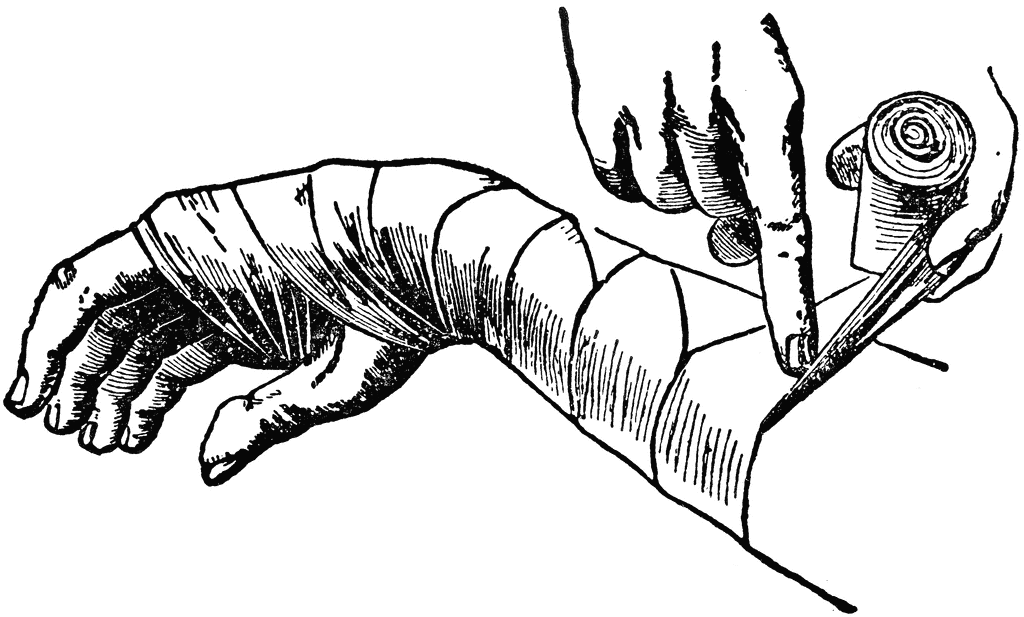
Most surgeons use a thin lining of stockinet or cellulose bandage to prevent
the plaster from sticking to the hairs and skin (Fig. 3.4). The use of a lining is
certainly recommended because it adds greatly to the comfort of the plaster. If
marked swelling is expected, as after an operation upon the limb, a more bulky
padding of surgical cotton wool should be used.
The plaster bandages are applied in two forms: round-and-round bandages
and longitudinal strips or ‘slabs’ to reinforce a particular area. Round-andround bandages must be applied smoothly without tension, the material being
drawn out to its full width at each turn. Slabs are prepared by unrolling a
bandage to and fro upon a table: an average slab consists of about 12 thicknesses. The slabs are placed at points of weakness or stress and are held in
place by further turns of plaster bandage.
A plaster is best dried simply by exposure to the air: artificial heating is
unnecessary. A plaster will not dry satisfactorily if it is kept covered by clothing
or bed-linen.
Synthetic (plastic) splinting materials are applied in much the same way as
plaster bandages, usually with warm water. Since they are stronger weight for
weight than plaster, fewer layers are required. Moulding to the body contours
is more difficult than with plaster bandages.


Immobilisation by internal fixation
Operative or internal fixation may be advised in the following circumstances:
1. to provide early control of limb fractures when conservative methods
would interfere with the management of other severe injuries, for instance
of the head, thorax or abdomen
2. as a method of choice in certain fractures, to secure immobilisation of the
fracture and to allow early mobility of the patient, e.g. in the elderly
patient with trochanteric hip fracture
3. when it has been necessary to operate upon a fracture to secure adequate
reduction
4. if it is impossible in a closed fracture to maintain an acceptable position by
splintage alone.
Methods of internal fixation. The following methods are currently in general
use (Fig. 3.16):
1. metal plate held by screws or locking plate (with screws fixed to the plate
by threaded holes)
2. intramedullary nail, with or without cross-screw fixation for locking
3. dynamic compression screw-plate
4. condylar screw-plate
5. tension band wiring
6. transfixion screws.
The choice of method depends upon the site and pattern of the fracture.
Plate and screws. This method is applicable to long bones. Usually a single
six-hole plate suffices, but an eight-hole plate may be preferred for larger bones.
Fixation by ordinary plates has the disadvantage that the bone fragments
are not forcibly pressed into close contact; indeed, if there is any absorption of the fracture surfaces the plate tends to hold the fragments apart, and this may
sometimes be a factor in the causation of delayed union. In order to counter this
disadvantage of simple plates and to improve coaptation at the time of plating,
special compression plates are available by which the fragments are forced
together before the plate is finally screwed home (compression plating).
Locking plate. A newer concept is the ‘locking plate’, that uses screws with
heads that are threaded and when tightened lock into matching threads in the
holes of the plate. This produces a more rigid fixation in terms of length and
angle, which is particularly valuable in comminuted fractures in osteoporotic
bone. It can also be inserted with less stripping of soft tissue that preserves
bone vascularity, particularly in the metaphyseal region.
Intramedullary nail. This technique is excellent for many fractures of the long
bones, especially when the fracture is near the middle of the shaft. It is used
regularly for fractures of the femur and tibia, and less commonly in the
humerus. The original Kuntscher-type nail designed for the femur was hollow
and of clover-leaf section and achieved fixation by its tight fit in the narrowest
isthmus of the shaft. This type has been replaced by the newer more versatile
locking nail with a rounder cross-section (Fig. 3.16), which offers notable
advantages. These have transverse holes at both ends, allowing the insertion of
transfixion (‘locking’) screws through bone and nail under image intensifier
radiographic control. This affords greater rigidity as well as resistance to
rotation forces allowing their use in comminuted fractures, particularly in the
wider medullary canal near the bone ends. A new design of thinner more flexible
solid nail is sometimes used for the management of shaft fractures in children.
Compression screw-plate. The compression screw-plate (dynamic hip screw) is
a standard method of fixation for fractures of the neck of the femur and for
trochanteric fractures (see Fig. 15.3). The screw component, which grips the
femoral head, slides telescopically in the barrel to allow the bone fragments to
be compressed together across the fracture. This compression effect is brought
about by tightening a screw in the base of the barrel.
Transfixion screws. The use of a transfixion screw has wide application in the
fixation of small detached fragments—for instance the capitulum of the
humerus, the olecranon process of the ulna or the medial malleolus of the tibia.
Kirschner wire fixation.  These thin flexible wires with sharpened ends are
available in a number of diameters and provide a useful alternative to
transfixion screws for the fixation of small bony fragments or for fractures of
the small bones in the hand and foot.
Tension band wiring. This technique of fixation is most commonly used in the
patella and olecranon, but can be applied to other small metaphyseal fragments
such as the medial malleolus. It uses the mechanical principle of converting the
tensile stresses of the muscles acting on the bone fragment, into a compressive
force at the fracture site. This is achieved by means of tightening an eccentric
figure-of-eight cerclage wire across the two fragments, stabilised by Kirschner
wires or a screw inserted at right angles to the fracture line (Fig. 3.16).

Metals for internal fixation
Metals used for internal fixation of fractures or for internal prostheses must be
resistant to corrosion in the tissues: silver, iron, ordinary steel and nickel-plated
steel are all unsuitable. A special stainless steel containing chromium, nickel
and molybdenum is widely used, but a non-ferrous alloy containing
chromium, cobalt and molybdenum has even better resistance to corrosion in
the body and is used for all types of internal appliance except wire, for whichit is technically unsuitable. The metallic element titanium and its alloys have
also proved resistant to corrosion in the body and are used increasingly for the
manufacture of prostheses and internal fixation devices.
The place of operative fixation
In recent years there has been an increasing use of internal fixation for the
treatment of limb fractures in most trauma centres, often as a deliberate first
choice. As will be seen in a later chapter, operative fixation is already accepted
as the best routine method of treating fractures of the neck and trochanteric
region of the femur in the elderly. Until recently, many fractures of the shafts of
the long bones have been treated conservatively—generally with excellent
results, although often at the cost of rather a long time in hospital or away from
work. The introduction of more sophisticated implants, inserted through small
incisions under radiological screening, and offering immediate fracture fixation,
has led to a dramatic change in this policy. In particular, intramedullary nailing
is now used for most fractures of the shaft of the femur or tibia.
The reasons for advocating surgical intervention for fractures that were
formerly managed conservatively are threefold. Firstly, there may be a
substantial reduction in the time that the patient must spend in hospital and
away from work. Secondly, in a favourable case function of the limb—and
particularly of the joints—may be restored earlier because the need for plaster
or other external splintage can often be eliminated. And thirdly, it is hoped that
by providing rigid fixation of the fracture, complications such as delayed union
and non-union will be reduced. In themselves, these objectives are unexceptionable, but there are arguments on the other side. The chief of these is that
operative fixation, especially when combined with open reduction, entails risks
that are absent or minimal with conservative treatment. Occasional fatalities—
for instance from pulmonary embolism—are probably unavoidable, and major
wound infection is by no means uncommon after lengthy open operations for
reduction and internal fixation. Extensive stripping of soft tissues from the
bone may also lead to adhesions that restrict joint movement, and may jeopardise the blood supply to the bone fragments, thereby hindering union. Thus
the objects of the operation may sometimes be defeated.
It is important to strike a fair balance between these conflicting arguments,
and to weigh up all the factors in every case: the age of the patient, the site and
nature of the fracture, problems of employment, and economic circumstances.
Advanced age should always weigh heavily in favour of an operation that will
enable the patient to get out of bed sooner, whereas anything that might favour
infection, such as an open wound or a pressure blister, should weigh heavily
against open operation. In such cases external fixation (p. 41) as distinct from
internal fixation has an important place.
The final decision on whether to use internal fixation or an alternative
conservative method of fracture treatment must be made by the surgeon. They
must be guided by their experience with the technique, the availability of the
implants, the operating and ward environments, and the incidence of wound
infection in other patients.


Ko Gemar LIKE ler.,xGemar leave COMMENT

0
Komen

Principles of fracture < Reduction >

This first principle must be qualified by the words ‘if necessary’. In many fractures reduction is unnecessary, either because there is no displacement or
because the displacement is immaterial to the final result. A considerable experience of fractures is needed before one can say with confidence
whether or not reduction is advisable in a given case. If it is judged that perfect
function can be restored without undue loss of time, despite some uncorrected
displacement of the fragments, there is clearly no object in striving for perfect
anatomical reduction. Indeed, meddlesome intervention may sometimes be
detrimental, especially if it entails open operation.
To take a simple example, there is no object in striving to replace perfectly
the broken fragments of a child’s clavicle, because normal function and
appearance will be restored without any intervention; the same applies to most fractures of the clavicle in adults. Likewise there is nothing to be gained instriving for perfect reduction of a fracture of the neck of the humerus in anelderly person—an ideal that may demand open operation for its attainment—
when good or better results may be expected from conservative treatmentdespite imperfect reduction.
In general, it may be said that imperfect apposition of the fragments can be
accepted much more readily than imperfect alignment (Fig. 3.1). For example,
in the shaft of the femur a loss of contact of half a diameter might be acceptable
whereas an angular deformity of 20º would usually demand an attempt at
improvement. When a joint surface is involved in a fracture, the articular
fragments must always be restored as nearly as possible to normal, to lessen
the risk of subsequent osteoarthritis.

METHODS OF REDUCTION
When reduction is decided upon it may be carried out in three ways:
1. by closed manipulation
2. by mechanical traction with or without manipulation
3. by open operation.
Manipulative reduction
Closed manipulation is the standard initial method of reducing most common
fractures. It is usually carried out under general anaesthesia, but local or
regional anaesthesia is sometimes appropriate. The technique is simply to
grasp the fragments through the soft tissues, to disimpact them if necessary,
and then to adjust them as nearly as possible to their correct position.
Reduction by mechanical traction
When the contraction of large muscles exerts a strong displacing force, some
mechanical aid may be necessary to draw the fragments out to the normal
length of the bone. This particularly applies to fractures of the shaft of the
femur, and to certain types of fracture or displacement of the cervical spine.
CHAPTER 3 31
Principles of fracture treatment
Fig. 3.1 Imperfect apposition (left) may
often be accepted, whereas malalignment
of more than a few degrees must usually
be corrected.
Often acceptable Seldom acceptable
Ch03-F10297.qxd  3/27/07  11:26 AM  Page 31Traction may be applied either by weights or by a screw device, and the aim
may be to gain full reduction rapidly at one sitting with anaesthesia, or to rely
upon gradual reduction by prolonged traction without anaesthesia.
Operative reduction
When an acceptable reduction cannot be obtained, or maintained, by these
conservative methods, the fragments are reduced under direct vision at open
operation. Open reduction may also be required for some fractures involving
articular surfaces, or when the fracture is complicated by damage to a nerve or
artery. When operative reduction is resorted to, the opportunity should always
be taken to fix the fragments internally to ensure that the position is maintained


Ko Gemar LIKE ler.,xGemar leave COMMENT

0
Komen

Principles of fracture < First aid >

The doctor who chances to be at the scene of an accident should seldom
attempt more than to ensure that the airway is clear, to control any external
haemorrhage, to cover any wound with a clean dressing, to provide some form
of immobilisation for a fractured limb, and to make the patient comfortable
while awaiting the arrival of the ambulance.
When it is necessary to move a patient with a long-bone fracture, it will be
found that pain is lessened if traction is applied to the limb while it is being
moved. If it is suspected that there may be a fracture of the spinal column,
special care is necessary in transport, lest injury to the spinal cord or cauda
equina be caused or aggravated. It is most important to avoid flexing the spine,
because flexion may cause or increase vertebral displacement, jeopardising the
spinal cord. In certain types of fracture, extension is also potentially dangerous
to the cord. Accordingly the patient should be lifted bodily on to a firm surface,
with care to avoid both flexion and extension. If a cervical collar is available, it
should be applied as a protection for the neck before moving the patient,
without allowing either flexion or extension of the neck during its application.
Temporary immobilisation for the long bones of the lower limb is
conveniently arranged by bandaging the two limbs together so that the sound
limb forms a splint for the injured one. In the upper limb, support may be
provided by bandaging the arm to the chest or, in the case of the forearm, by
improvising a sling.
Haemorrhage hardly ever demands a tourniquet for its control. All ordinary
bleeding can be controlled adequately by firm bandaging over a pad. Only if
profuse pulsatile (arterial) bleeding persists despite firm pressure over the
wound, with the patient recumbent, does the need for a tourniquet arise.
Pending its application, firm manual pressure over the main artery at the root
of the limb may be applied to control the bleeding. If a tourniquet is applied,
those attending the patient should be made aware of the fact and of the time of
its application. If necessary, a note to this effect should be sent with the patient
to ensure that the tourniquet is not inadvertently left in place for too long.
Ch03-F10297.qxd  3/27/07  11:26 AM  Page 29If morphine or a similar drug is given at the scene of the accident a note to
that effect should be sent with the patient on admission to hospital.
Clinical assessment
It must be emphasised again that an immediate assessment of the whole
patient is required to exclude injuries to other systems before examination of
the skeletal injury. Examination of the limb should determine:
1. whether there is a wound communicating with the fracture
2. whether there is evidence of a vascular injury
3. whether there is evidence of a nerve injury
4. whether there is evidence of visceral injury.
Resuscitation
Many patients with severe or multiple fractures, or fractures associated with
other visceral injuries, are shocked on arrival at hospital. Time must be spent
on resuscitation and dealing with any other life-threatening injuries before
definitive treatment for the fracture is begun. Haemorrhagic shock can develop
rapidly when there has been a rapid loss of a large volume of blood. The mainstay of treatment is the immediate replenishment of the circulating blood volume,
either with transfused blood when time permits cross-matching, or alternatively
by the use of plasma expanders and blood substitutes. Electrolytes, such as
isotonic saline or Rimmer’s lactate solution, can be used to establish
intravenous infusion but are of little value in replacing lost blood. Colloid
solutions which remain within the circulation are of more value and include
dextran, a high-molecular-weight polysaccharide, gelatin solution derived
from animal protein, or a plasma protein fraction solution of human albumin
with a small proportion of globulin. Transfusion with colloids or whole blood
is usually only required in patients with blood loss greater than 1 litre.


Ko Gemar LIKE ler.,xGemar leave COMMENT

Friday 9 September 2011

0
Komen

Perang Dunia Ke-2

Assalammuailaikum semua..,
wahhh..,lama rasa nyer ktew x bersua eak..??? bkn nyer x bersemua lngsung..,tpi aq yg xde wat entry bru kan???mesti yg mne singgah..,mesti bosan yg teramat kan..???? yela..,kat blog aq nei bkn nyer de pew yg menarik pun..,hahhaha..,xpew..,aq faham..,

okey tujuan aq bersandar kat cne ary nei adalah nak luah kan rasa aty yg terbuku n kegeraman jiwa rohani aq..,hahhah..,nei aq nak crte kan..,tpi kan..???tbe2 jew aq cam malas nak menaip..,aduhh..,tkot t aq crte..,terbakar lak tempat nei..,hahhaha..,kisah nyer sample jew..,td aq gduh blek ngan senior2 ku..hahhaha..,''KO SAPA HAHH!!! BP UMOR KO HAHH!!..???'' mu gat mu ckp ngan bdk skolah kew???sowi ler weiii..,i remaja k..,adult k..,suka aty aq ler..,ko jgn gat..,aq dok m'galah..,tanda aq kalah..,hahhaha..,sowi ler wei..,TERPAKSA jew..!!! ckit pun aq xkan maaf kan kowg..,ha'ahhh!!!!!!! aq pedulli pew..,hak aq..,uuhh!!

Klu dah tau dri tu leader..,arrange ler umah tu ngan bgus..,jgn gat ko leader..,suka aty mak tiri ko nak bawak jantan masuk umah..,!!!! nak bwk kerabat2 ko msuk..???weeiii..,sowi ler eak..,yg salah ttp salah..,nak wat pew aq m'galah..,!! klu setakat ckp dri tu leader..,smua owg leh ckp lew weii..,!!! smua owg leh m'gaku ler siott..,!!!tapi leader yg bnor2 okey..,dpt arrange umah ngan btol n t'atur..,xgne pggn title ''LEADER'',tpi tgus langsung..,!!! nak kata xde mata.,ada..,leh sihat melihat lg..,gne ler pacarindera tu utk perkara yg baek..,aduhyaiii!!!!..,

okey ler kowg..,aq nak out nei..,battery pun dah nyawa2 ikan nei..,so..,nati de masa temu lg k???daaa..,


Ko Gemar LIKE ler.,xGemar leave COMMENT

Monday 5 September 2011

0
Komen

Normal Range..,

HEMATOLOGY – Red Blood Cells
RBC (Male) 4.2 – 5.6 10^6 / µL [Scientific Notation: 10^6 = 1,000,000]
RBC (Female) 3.8 – 5.1 10^6 / µL
RBC (Child) 3.5 – 5.0 10^6 / µL
HEMATOLOGY – White Blood Cells
WBC (Male) 3.8 – 11.0 10^3 / mm3 [Scientific Notation: 10^3 = 1,000]
WBC (Female) 3.8 – 11.0 10^3 / mm3
WBC (Child) 5.0 – 10.0 10^3 / mm3
HEMOGLOBIN
Hgb (Male) 14 – 18 g/dL
Hgb (Female) 11 – 16 g/dL
Hgb (Child) 10 – 14 g/dL
Hgb (Newborn) 15 – 25 g/dL
HEMATOCRIT
Hct (Male) 39 – 54%
Hct (Female) 34 – 47%
Hct (Child) 30 – 42%
MCV 78 – 98 fL
MCH 27 – 35 pg
MCHC 31 – 37%
neutrophils 50 – 81%
bands 1 – 5%
lymphocytes 14 – 44%
monocytes 2 – 6%
eosinophils 1 – 5%
basophils 0 – 1%
CARDIAC MARKERS
troponin I 0 – 0.1 ng/ml (onset: 4-6 hrs, peak:
12-24 hrs, return to normal: 4-7 days)
troponin T 0 – 0.2 ng/ml (onset: 3-4 hrs, peak:
10-24 hrs, return to normal: 10-14 days)
myoglobin (Male) 10 – 95 ng/ml (onset: 1-3 hrs, peak:
6-10 hrs, return to normal: 12-24 hrs)
myoglobin (Female) 10 – 65 ng/ml (onset: 1-3 hrs, peak:
6-10 hrs, return to normal: 12-24 hrs)
GENERAL CHEMISTRY
acetone 0.3 – 2.0 mg%
albumin 3.5 – 5.0 gm/dL
alkaline phosphatase 32 – 110 U/L
anion gap 5 – 16 mEq/L
ammonia 11 – 35 µmol/L
amylase 50 – 150 U/dL
AST,SGOT (Male) 7 – 21 U/L
AST,SGOT (Female) 6 – 18 U/L
bilirubin, direct 0.0 – 0.4 mg/dL
bilirubin, indirect total minus direct
bilirubin, total 0.2 – 1.4 mg/dL
BUN 6 – 23 mg/dL
calcium (total) 8 – 11 mg/dL
carbon dioxide 21 – 34 mEq/L
carbon monoxide symptoms at greater than or equal to 10% saturation
chloride 96 – 112 mEq/L
creatine (Male) 0.2 – 0.6 mg/dL
creatine (Female) 0.6 – 1.0 mg/dL
creatinine 0.6 – 1.5 mg/dL
ethanol 0 mg%; Coma:
greater than or equal to 400 – 500 mg%
folic acid 2.0 – 21 ng/mL
glucose 65 – 99 mg/dL
(diuresis greater than or equal to 180 mg/dL)
HDL (Male) 25 – 65 mg/dL
HDL (Female) 38 – 94 mg/dL
iron 52 – 169 µg/dL
iron binding capacity 246 – 455 µg/dL
lactic acid 0.4 – 2.3 mEq/L
lactate 0.3 – 2.3 mEq/L
lipase 10 – 140 U/L
magnesium 1.5 – 2.5 mg/dL
osmolarity 276 – 295 mOsm/kg
parathyroid hormone 12 – 68 pg/mL
phosphorus 2.2 – 4.8 mg/dL
potassium 3.5 – 5.5 mEq/L
SGPT 8 – 32 U/L
sodium 135 – 148 mEq/L
T3 0.8 – 1.1 µg/dL
thyroglobulin less than 55 ng/mL
thyroxine (T4) (total) 5 – 13 µg/dL
total protein 5 – 9 gm/dL
TSH Less than 9 µU/mL
urea nitrogen 8 – 25 mg/dL
uric acid (Male) 3.5 – 7.7 mg/dL
uric acid (Female) 2.5 – 6.6 mg/dL
LIPID PANEL (Adult)
cholesterol (total) Less than 200 mg/dL desirable
cholesterol (HDL) 30 – 75 mg/dL
cholesterol (LDL) Less than 130 mg/dL desirable
triglycerides (Male) Greater than 40 – 170 mg/dL
triglycerides (Female) Greater than 35 – 135 mg/dL
URINE
color Straw
specific gravity 1.003 – 1.040
pH 4.6 – 8.0
Na 10 – 40 mEq/L
K Less than 8 mEq/L
C1 Less than 8 mEq/L
protein 1 – 15 mg/dL
osmolality 80 – 1300 mOsm/L
24 HOUR URINE
amylase 250 – 1100 IU / 24 hr
calcium 100 – 250 mg / 24 hr
chloride 110 – 250 mEq / 24 hr
creatinine 1 – 2 g / 24 hr
creatine clearance (Male) 100 – 140 mL / min
creatine clearance (Male) 16 – 26 mg / kg / 24 hr
creatine clearance (Female) 80 – 130 mL / min
creatine clearance (Female) 10 – 20 mg / kg / 24 hr
magnesium 6 – 9 mEq / 24 hr
osmolality 450 – 900 mOsm / kg
phosphorus 0.9 – 1.3 g / 24 hr
potassium 35 – 85 mEq / 24 hr
protein 0 – 150 mg / 24 hr
sodium 30 – 280 mEq / 24 hr
urea nitrogen 10 – 22 gm / 24 hr
uric acid 240 – 755 mg / 24 hr
COAGULATION
ACT 90 – 130 seconds
APTT 21 – 35 seconds
platelets 140,000 – 450,000 /ml
plasminogen 62 – 130%
PT 10 – 14 seconds
PTT 32 – 45 seconds
FSP Less than 10 µg/dL
fibrinogen 160 – 450 mg/dL
bleeding time 3 – 7 minutes
thrombin time 11 – 15 seconds
CEREBRAL SPINAL FLUID
appearance clear
glucose 40 – 85 mg/dL
osmolality 290 – 298 mOsm/L
pressure 70 – 180 mm/H2O
protein 15 – 45 mg/dL
total cell count 0 – 5 cells
WBCs 0 – 6 / µL
HEMODYNAMIC PARAMETERS
cardiac index 2.5 – 4.2 L / min / m2
cardiac output 4 – 8 LPM
left ventricular stroke work index 40 – 70 g / m2 / beat
right ventricular stroke work index 7 – 12 g / m2 / beat
mean arterial pressure 70 – 105 mm Hg
pulmonary vascular resistance 155 – 255 dynes / sec / cm to the negative 5
pulmonary vascular resistance index 255 – 285 dynes / sec / cm to the negative 5
stroke volume 60 – 100 mL / beat
stroke volume index 40 – 85 mL / m2 / beat
systemic vascular resistance 900 – 1600 dynes / sec / cm to the negative 5
systemic vascular resistance index 1970 – 2390 dynes / sec / cm to the negative 5
systolic arterial pressure 90 – 140 mm Hg
diastolic arterial pressure 60 – 90 mm Hg
central venous pressure 2 – 6 mm Hg; 2.5 – 12 cm H2O
ejection fraction 60 – 75%
left arterial pressure 4 – 12 mm Hg
right atrial pressure 4 – 6 mm Hg
pulmonary artery systolic 15 – 30 mm Hg
pulmonary artery diastolic 5 – 15 mm Hg
pulmonary artery pressure 10 – 20 mm Hg
pulmonary artery wedge pressure 4 – 12 mm Hg
pulmonary artery end diastolic pressure 8 – 10 mm Hg
right ventricular end diastolic pressure 0 – 8 mm Hg
NEUROLOGICAL VALUES
cerebral perfusion pressure 70 – 90 mm Hg
intracranial pressure 5 – 15 mm Hg or 5 – 10 cm H2O
ARTERIAL VALUES
pH 7.35 – 7.45
PaCO2 35 – 45 mm Hg
HCO3 22 – 26 mEq/L
O2 saturation 96 – 100%
PaO2 85 – 100 mm Hg
BE -2 to +2 mmol/L
VENOUS VALUES
pH 7.31 – 7.41
PaCO2 41 – 51 mm Hg
HCO3 22 – 29 mEq/L
O2 saturation 60 – 85%
PaO2 30 – 40 mm Hg
BE 0 to +4 mmol/L


Ko Gemar LIKE ler.,xGemar leave COMMENT