ayat gerak

Aku dye..cinta kami..kisah kami..sayang dye..bila kowg dah masuk blog kami..kowg kena gak follow kami tau!!! ^_^

CoR3t@n Kami...,

ASSALAMMUAILAIKUM....
selamat datang kew ruangan kami..,so..,d sbb kn nei ruangan kami..,suka aty kami ler nak tulis entry pew..,n nak letak pew..,klu rasa x suka..,wat xtau jew eak..,i don't care about that!!!!!!! cbb ruangan nei sentiasa nyer ada pic d setiap warkah..,k ler..,hope ur all enjoy k..dah singgah jgn lupa follow n leave ur comment.,,daaa^_^

Sunday, 11 September 2011

0
Komen

Principles of fracture < Rehabilitation >

Improved results in the treatment of fractures owe much to rehabilitation, perhaps the most important of the three great principles of fracture treatment. Reduction is often unnecessary; immobilisation is often unnecessary; rehabilitation is always essential. In Britain, much of the credit for early enlightenment on the
principles of rehabilitation must go to Watson-Jones.
Rehabilitation should begin as soon as the fracture comes under definitive
treatment. Its purpose is twofold: first, to preserve function so far as possible
while the fracture is uniting and second, to restore function to normal when the
fracture is united. This purpose is achieved not so much by any passive treatment as by encouraging patients to help themselves.
The two essential methods of rehabilitation are active use and active exercises. Except in cases of minor injury, the patient should, ideally, be under the
supervision of a physiotherapist throughout the whole duration of treatment.

Active use
This implies that the patient must continue to use the injured part as naturally
as possible within the limitations imposed by necessary treatment (Fig. 3.17).
The degree of function that can be retained depends upon the nature of the
fracture, the risk of redisplacement of the fragments, and the extent of any
necessary splintage. Although in some injuries rest may be necessary in the
early days or weeks, there should be a graduated return to activity as soon as
it can be allowed without risk.

Active exercises
These comprise exercises for the muscles and joints. They should be
encouraged from an early stage. While a limb is immobilised in a plaster or
splint, exercises must be directed mainly to the preservation of muscle function
by static contractions. The ability to contract a muscle without moving a joint
is soon acquired under proper supervision.
When restrictive splints are no longer required, exercises should be directed
to mobilising the joints and building up the power of the muscles. Finally,
when the fracture is soundly united, treatment may be intensified, movementsbeing carried out against gradually increased resistance until normal power is
regained.
Although every adult patient with a major fracture should attend for
supervised exercises as often as possible, it should be impressed upon the
patient that this organised treatment plays only a part in the rehabilitation, and
that much—indeed most—depends upon continuing normal activities so far as
possible when the patient is away from the department. Physiotherapy is often
enormously helpful, but it should supplement, not supplant, the patient’s own
independent efforts (Figs 3.18 & 3.19).So far as children are concerned, supervised exercises are relatively
unimportant, and in most cases children may safely be left to their own
endeavours, aided when necessary by encouragement from the parents, who
should always be fully informed of the programme of treatment and the likely
course of events.
Continuous passive motion
In the knowledge that movement between joint surfaces favours the preservation of healthy articular cartilage, surgeons and biomechanical engineers
have designed machines that provide continuous to-and-fro movement at a
joint without any effort on the part of the patient. The range of movement can
be varied as required, being increased gradually as the joint becomes more
mobile. This technique of exercising joints passively has many applications: it
is particularly valuable in situations where restriction of mobility tends to be
hard to overcome, for instance in the knee after fracture of the femoral shaft or
after the operation of quadricepsplasty.


Ko Gemar LIKE ler.,xGemar leave COMMENT

0
Komen

Principles of fracture < immobilisation >

Like reduction, this second great principle of fracture treatment must be
qualified by the words ‘if necessary’. Whereas some fractures must be splinted
rigidly, many do not require immobilisation to ensure union, and excessive
immobilisation is actually harmful in some (Figs 3.2 & 3.3).

INDICATIONS FOR IMMOBILISATION
There are only three reasons for immobilising a fracture:
1. to prevent displacement or angulation of the fragments
2. to prevent movement that might interfere with union
3. to relieve pain.


Prevention of displacement or angulation
As a general rule, the broken fragments will not become displaced more
severely than they were at the time of the original injury. Therefore, if the
original position is acceptable, immobilisation to prevent further displacement
is often unnecessary. In fractures of the shafts of the major long bones, however,
immobilisation is usually necessary in order to maintain correct alignment.

Prevention of movement
As has been mentioned already, absolute immobility is not always essential to
union of a fracture. It is only when movement might shear the delicate capillaries bridging the fracture that it is undesirable, and, theoretically, rotation
movements are worst in this respect. There are three fractures that constantly
demand immobilisation to ensure their union—namely, those of the scaphoid
bone, of the shaft of the ulna, and of the neck of the femur.
Examples of fractures that heal well without immobilisation are those of
the ribs, clavicle and scapula, and stable fractures of the pelvic ring.
Immobilisation is also unnecessary for certain fractures of the humerus and
femur, and many fractures of the metacarpals, metatarsals and phalanges. In
some fractures, excessive immobilisation may do more harm than good. The
injured hand, in particular, tolerates prolonged immobilisation badly. Whereas
the wrist may be immobilised for many weeks or even months with impunity,
to immobilise injured fingers for a long time is to court disaster in the form of
permanent joint stiffness.

Relief of pain
Probably in about half of all the cases in which a fracture is immobilised the
main reason for immobilisation is to relieve pain. With the limb thus made
comfortable, it can be used much more effectively than would otherwise be
possible.

METHODS OF IMMOBILISATION
When immobilisation is deemed necessary there are four methods by which it
may be effected:
1. by a plaster of Paris cast or other external splint
2. by continuous traction
3. by external fixation
4. by internal fixation.

Immobilisation by plaster, splint or brace
For most fractures the standard method of immobilisation is by a plaster of
Paris cast. Also available are various proprietary substitutes for plaster, which
offer the advantages of lighter weight, radiolucency and imperviousness to
water, though at much greater cost. Most such products are also more difficult
to apply; nevertheless, they are being used on an increasing scale. For some
fractures a splint made from metal, wood or plastic is more appropriate—for
example, the Thomas’s splint for fractures of the shaft of the femur, or a plastic
collar for certain injuries of the cervical spine.
Plaster technique. Plaster of Paris is hemihydrated calcium sulphate. It reacts
with water to form hydrated calcium sulphate. The reaction is exothermic, a
fact that is evidenced by noticeable warming of the plaster during setting.
Plaster bandages may be prepared by impregnating rolls of book muslin
with the dry powdered plaster, but except in a few developing countries, most
hospitals now use ready-made proprietary bandages. These are best used with
cold water because setting is too rapid with warm water.
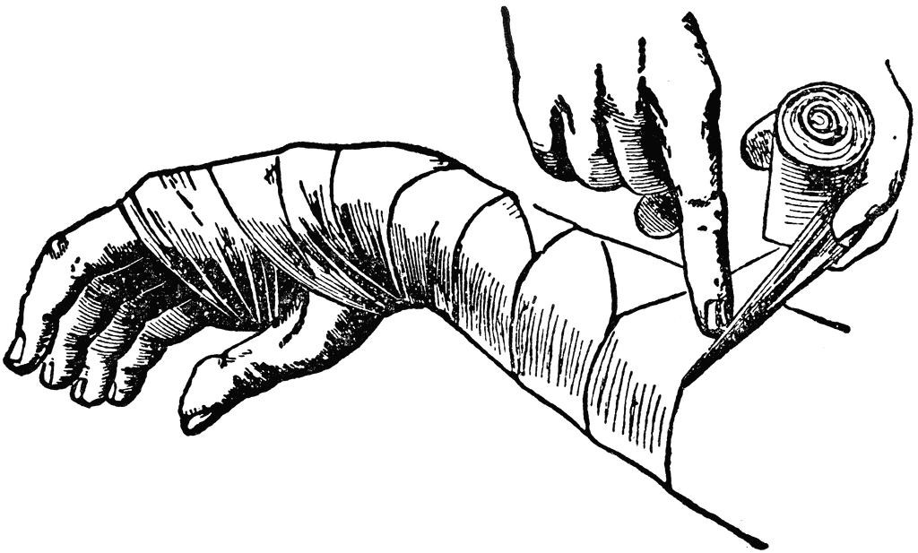
Most surgeons use a thin lining of stockinet or cellulose bandage to prevent
the plaster from sticking to the hairs and skin (Fig. 3.4). The use of a lining is
certainly recommended because it adds greatly to the comfort of the plaster. If
marked swelling is expected, as after an operation upon the limb, a more bulky
padding of surgical cotton wool should be used.
The plaster bandages are applied in two forms: round-and-round bandages
and longitudinal strips or ‘slabs’ to reinforce a particular area. Round-andround bandages must be applied smoothly without tension, the material being
drawn out to its full width at each turn. Slabs are prepared by unrolling a
bandage to and fro upon a table: an average slab consists of about 12 thicknesses. The slabs are placed at points of weakness or stress and are held in
place by further turns of plaster bandage.
A plaster is best dried simply by exposure to the air: artificial heating is
unnecessary. A plaster will not dry satisfactorily if it is kept covered by clothing
or bed-linen.
Synthetic (plastic) splinting materials are applied in much the same way as
plaster bandages, usually with warm water. Since they are stronger weight for
weight than plaster, fewer layers are required. Moulding to the body contours
is more difficult than with plaster bandages.


Immobilisation by internal fixation
Operative or internal fixation may be advised in the following circumstances:
1. to provide early control of limb fractures when conservative methods
would interfere with the management of other severe injuries, for instance
of the head, thorax or abdomen
2. as a method of choice in certain fractures, to secure immobilisation of the
fracture and to allow early mobility of the patient, e.g. in the elderly
patient with trochanteric hip fracture
3. when it has been necessary to operate upon a fracture to secure adequate
reduction
4. if it is impossible in a closed fracture to maintain an acceptable position by
splintage alone.
Methods of internal fixation. The following methods are currently in general
use (Fig. 3.16):
1. metal plate held by screws or locking plate (with screws fixed to the plate
by threaded holes)
2. intramedullary nail, with or without cross-screw fixation for locking
3. dynamic compression screw-plate
4. condylar screw-plate
5. tension band wiring
6. transfixion screws.
The choice of method depends upon the site and pattern of the fracture.
Plate and screws. This method is applicable to long bones. Usually a single
six-hole plate suffices, but an eight-hole plate may be preferred for larger bones.
Fixation by ordinary plates has the disadvantage that the bone fragments
are not forcibly pressed into close contact; indeed, if there is any absorption of the fracture surfaces the plate tends to hold the fragments apart, and this may
sometimes be a factor in the causation of delayed union. In order to counter this
disadvantage of simple plates and to improve coaptation at the time of plating,
special compression plates are available by which the fragments are forced
together before the plate is finally screwed home (compression plating).
Locking plate. A newer concept is the ‘locking plate’, that uses screws with
heads that are threaded and when tightened lock into matching threads in the
holes of the plate. This produces a more rigid fixation in terms of length and
angle, which is particularly valuable in comminuted fractures in osteoporotic
bone. It can also be inserted with less stripping of soft tissue that preserves
bone vascularity, particularly in the metaphyseal region.
Intramedullary nail. This technique is excellent for many fractures of the long
bones, especially when the fracture is near the middle of the shaft. It is used
regularly for fractures of the femur and tibia, and less commonly in the
humerus. The original Kuntscher-type nail designed for the femur was hollow
and of clover-leaf section and achieved fixation by its tight fit in the narrowest
isthmus of the shaft. This type has been replaced by the newer more versatile
locking nail with a rounder cross-section (Fig. 3.16), which offers notable
advantages. These have transverse holes at both ends, allowing the insertion of
transfixion (‘locking’) screws through bone and nail under image intensifier
radiographic control. This affords greater rigidity as well as resistance to
rotation forces allowing their use in comminuted fractures, particularly in the
wider medullary canal near the bone ends. A new design of thinner more flexible
solid nail is sometimes used for the management of shaft fractures in children.
Compression screw-plate. The compression screw-plate (dynamic hip screw) is
a standard method of fixation for fractures of the neck of the femur and for
trochanteric fractures (see Fig. 15.3). The screw component, which grips the
femoral head, slides telescopically in the barrel to allow the bone fragments to
be compressed together across the fracture. This compression effect is brought
about by tightening a screw in the base of the barrel.
Transfixion screws. The use of a transfixion screw has wide application in the
fixation of small detached fragments—for instance the capitulum of the
humerus, the olecranon process of the ulna or the medial malleolus of the tibia.
Kirschner wire fixation.  These thin flexible wires with sharpened ends are
available in a number of diameters and provide a useful alternative to
transfixion screws for the fixation of small bony fragments or for fractures of
the small bones in the hand and foot.
Tension band wiring. This technique of fixation is most commonly used in the
patella and olecranon, but can be applied to other small metaphyseal fragments
such as the medial malleolus. It uses the mechanical principle of converting the
tensile stresses of the muscles acting on the bone fragment, into a compressive
force at the fracture site. This is achieved by means of tightening an eccentric
figure-of-eight cerclage wire across the two fragments, stabilised by Kirschner
wires or a screw inserted at right angles to the fracture line (Fig. 3.16).

Metals for internal fixation
Metals used for internal fixation of fractures or for internal prostheses must be
resistant to corrosion in the tissues: silver, iron, ordinary steel and nickel-plated
steel are all unsuitable. A special stainless steel containing chromium, nickel
and molybdenum is widely used, but a non-ferrous alloy containing
chromium, cobalt and molybdenum has even better resistance to corrosion in
the body and is used for all types of internal appliance except wire, for whichit is technically unsuitable. The metallic element titanium and its alloys have
also proved resistant to corrosion in the body and are used increasingly for the
manufacture of prostheses and internal fixation devices.
The place of operative fixation
In recent years there has been an increasing use of internal fixation for the
treatment of limb fractures in most trauma centres, often as a deliberate first
choice. As will be seen in a later chapter, operative fixation is already accepted
as the best routine method of treating fractures of the neck and trochanteric
region of the femur in the elderly. Until recently, many fractures of the shafts of
the long bones have been treated conservatively—generally with excellent
results, although often at the cost of rather a long time in hospital or away from
work. The introduction of more sophisticated implants, inserted through small
incisions under radiological screening, and offering immediate fracture fixation,
has led to a dramatic change in this policy. In particular, intramedullary nailing
is now used for most fractures of the shaft of the femur or tibia.
The reasons for advocating surgical intervention for fractures that were
formerly managed conservatively are threefold. Firstly, there may be a
substantial reduction in the time that the patient must spend in hospital and
away from work. Secondly, in a favourable case function of the limb—and
particularly of the joints—may be restored earlier because the need for plaster
or other external splintage can often be eliminated. And thirdly, it is hoped that
by providing rigid fixation of the fracture, complications such as delayed union
and non-union will be reduced. In themselves, these objectives are unexceptionable, but there are arguments on the other side. The chief of these is that
operative fixation, especially when combined with open reduction, entails risks
that are absent or minimal with conservative treatment. Occasional fatalities—
for instance from pulmonary embolism—are probably unavoidable, and major
wound infection is by no means uncommon after lengthy open operations for
reduction and internal fixation. Extensive stripping of soft tissues from the
bone may also lead to adhesions that restrict joint movement, and may jeopardise the blood supply to the bone fragments, thereby hindering union. Thus
the objects of the operation may sometimes be defeated.
It is important to strike a fair balance between these conflicting arguments,
and to weigh up all the factors in every case: the age of the patient, the site and
nature of the fracture, problems of employment, and economic circumstances.
Advanced age should always weigh heavily in favour of an operation that will
enable the patient to get out of bed sooner, whereas anything that might favour
infection, such as an open wound or a pressure blister, should weigh heavily
against open operation. In such cases external fixation (p. 41) as distinct from
internal fixation has an important place.
The final decision on whether to use internal fixation or an alternative
conservative method of fracture treatment must be made by the surgeon. They
must be guided by their experience with the technique, the availability of the
implants, the operating and ward environments, and the incidence of wound
infection in other patients.


Ko Gemar LIKE ler.,xGemar leave COMMENT

0
Komen

Principles of fracture < Reduction >

This first principle must be qualified by the words ‘if necessary’. In many fractures reduction is unnecessary, either because there is no displacement or
because the displacement is immaterial to the final result. A considerable experience of fractures is needed before one can say with confidence
whether or not reduction is advisable in a given case. If it is judged that perfect
function can be restored without undue loss of time, despite some uncorrected
displacement of the fragments, there is clearly no object in striving for perfect
anatomical reduction. Indeed, meddlesome intervention may sometimes be
detrimental, especially if it entails open operation.
To take a simple example, there is no object in striving to replace perfectly
the broken fragments of a child’s clavicle, because normal function and
appearance will be restored without any intervention; the same applies to most fractures of the clavicle in adults. Likewise there is nothing to be gained instriving for perfect reduction of a fracture of the neck of the humerus in anelderly person—an ideal that may demand open operation for its attainment—
when good or better results may be expected from conservative treatmentdespite imperfect reduction.
In general, it may be said that imperfect apposition of the fragments can be
accepted much more readily than imperfect alignment (Fig. 3.1). For example,
in the shaft of the femur a loss of contact of half a diameter might be acceptable
whereas an angular deformity of 20º would usually demand an attempt at
improvement. When a joint surface is involved in a fracture, the articular
fragments must always be restored as nearly as possible to normal, to lessen
the risk of subsequent osteoarthritis.

METHODS OF REDUCTION
When reduction is decided upon it may be carried out in three ways:
1. by closed manipulation
2. by mechanical traction with or without manipulation
3. by open operation.
Manipulative reduction
Closed manipulation is the standard initial method of reducing most common
fractures. It is usually carried out under general anaesthesia, but local or
regional anaesthesia is sometimes appropriate. The technique is simply to
grasp the fragments through the soft tissues, to disimpact them if necessary,
and then to adjust them as nearly as possible to their correct position.
Reduction by mechanical traction
When the contraction of large muscles exerts a strong displacing force, some
mechanical aid may be necessary to draw the fragments out to the normal
length of the bone. This particularly applies to fractures of the shaft of the
femur, and to certain types of fracture or displacement of the cervical spine.
CHAPTER 3 31
Principles of fracture treatment
Fig. 3.1 Imperfect apposition (left) may
often be accepted, whereas malalignment
of more than a few degrees must usually
be corrected.
Often acceptable Seldom acceptable
Ch03-F10297.qxd  3/27/07  11:26 AM  Page 31Traction may be applied either by weights or by a screw device, and the aim
may be to gain full reduction rapidly at one sitting with anaesthesia, or to rely
upon gradual reduction by prolonged traction without anaesthesia.
Operative reduction
When an acceptable reduction cannot be obtained, or maintained, by these
conservative methods, the fragments are reduced under direct vision at open
operation. Open reduction may also be required for some fractures involving
articular surfaces, or when the fracture is complicated by damage to a nerve or
artery. When operative reduction is resorted to, the opportunity should always
be taken to fix the fragments internally to ensure that the position is maintained


Ko Gemar LIKE ler.,xGemar leave COMMENT

0
Komen

Principles of fracture < First aid >

The doctor who chances to be at the scene of an accident should seldom
attempt more than to ensure that the airway is clear, to control any external
haemorrhage, to cover any wound with a clean dressing, to provide some form
of immobilisation for a fractured limb, and to make the patient comfortable
while awaiting the arrival of the ambulance.
When it is necessary to move a patient with a long-bone fracture, it will be
found that pain is lessened if traction is applied to the limb while it is being
moved. If it is suspected that there may be a fracture of the spinal column,
special care is necessary in transport, lest injury to the spinal cord or cauda
equina be caused or aggravated. It is most important to avoid flexing the spine,
because flexion may cause or increase vertebral displacement, jeopardising the
spinal cord. In certain types of fracture, extension is also potentially dangerous
to the cord. Accordingly the patient should be lifted bodily on to a firm surface,
with care to avoid both flexion and extension. If a cervical collar is available, it
should be applied as a protection for the neck before moving the patient,
without allowing either flexion or extension of the neck during its application.
Temporary immobilisation for the long bones of the lower limb is
conveniently arranged by bandaging the two limbs together so that the sound
limb forms a splint for the injured one. In the upper limb, support may be
provided by bandaging the arm to the chest or, in the case of the forearm, by
improvising a sling.
Haemorrhage hardly ever demands a tourniquet for its control. All ordinary
bleeding can be controlled adequately by firm bandaging over a pad. Only if
profuse pulsatile (arterial) bleeding persists despite firm pressure over the
wound, with the patient recumbent, does the need for a tourniquet arise.
Pending its application, firm manual pressure over the main artery at the root
of the limb may be applied to control the bleeding. If a tourniquet is applied,
those attending the patient should be made aware of the fact and of the time of
its application. If necessary, a note to this effect should be sent with the patient
to ensure that the tourniquet is not inadvertently left in place for too long.
Ch03-F10297.qxd  3/27/07  11:26 AM  Page 29If morphine or a similar drug is given at the scene of the accident a note to
that effect should be sent with the patient on admission to hospital.
Clinical assessment
It must be emphasised again that an immediate assessment of the whole
patient is required to exclude injuries to other systems before examination of
the skeletal injury. Examination of the limb should determine:
1. whether there is a wound communicating with the fracture
2. whether there is evidence of a vascular injury
3. whether there is evidence of a nerve injury
4. whether there is evidence of visceral injury.
Resuscitation
Many patients with severe or multiple fractures, or fractures associated with
other visceral injuries, are shocked on arrival at hospital. Time must be spent
on resuscitation and dealing with any other life-threatening injuries before
definitive treatment for the fracture is begun. Haemorrhagic shock can develop
rapidly when there has been a rapid loss of a large volume of blood. The mainstay of treatment is the immediate replenishment of the circulating blood volume,
either with transfused blood when time permits cross-matching, or alternatively
by the use of plasma expanders and blood substitutes. Electrolytes, such as
isotonic saline or Rimmer’s lactate solution, can be used to establish
intravenous infusion but are of little value in replacing lost blood. Colloid
solutions which remain within the circulation are of more value and include
dextran, a high-molecular-weight polysaccharide, gelatin solution derived
from animal protein, or a plasma protein fraction solution of human albumin
with a small proportion of globulin. Transfusion with colloids or whole blood
is usually only required in patients with blood loss greater than 1 litre.


Ko Gemar LIKE ler.,xGemar leave COMMENT

Friday, 9 September 2011

0
Komen

Perang Dunia Ke-2

Assalammuailaikum semua..,
wahhh..,lama rasa nyer ktew x bersua eak..??? bkn nyer x bersemua lngsung..,tpi aq yg xde wat entry bru kan???mesti yg mne singgah..,mesti bosan yg teramat kan..???? yela..,kat blog aq nei bkn nyer de pew yg menarik pun..,hahhaha..,xpew..,aq faham..,

okey tujuan aq bersandar kat cne ary nei adalah nak luah kan rasa aty yg terbuku n kegeraman jiwa rohani aq..,hahhah..,nei aq nak crte kan..,tpi kan..???tbe2 jew aq cam malas nak menaip..,aduhh..,tkot t aq crte..,terbakar lak tempat nei..,hahhaha..,kisah nyer sample jew..,td aq gduh blek ngan senior2 ku..hahhaha..,''KO SAPA HAHH!!! BP UMOR KO HAHH!!..???'' mu gat mu ckp ngan bdk skolah kew???sowi ler weiii..,i remaja k..,adult k..,suka aty aq ler..,ko jgn gat..,aq dok m'galah..,tanda aq kalah..,hahhaha..,sowi ler wei..,TERPAKSA jew..!!! ckit pun aq xkan maaf kan kowg..,ha'ahhh!!!!!!! aq pedulli pew..,hak aq..,uuhh!!

Klu dah tau dri tu leader..,arrange ler umah tu ngan bgus..,jgn gat ko leader..,suka aty mak tiri ko nak bawak jantan masuk umah..,!!!! nak bwk kerabat2 ko msuk..???weeiii..,sowi ler eak..,yg salah ttp salah..,nak wat pew aq m'galah..,!! klu setakat ckp dri tu leader..,smua owg leh ckp lew weii..,!!! smua owg leh m'gaku ler siott..,!!!tapi leader yg bnor2 okey..,dpt arrange umah ngan btol n t'atur..,xgne pggn title ''LEADER'',tpi tgus langsung..,!!! nak kata xde mata.,ada..,leh sihat melihat lg..,gne ler pacarindera tu utk perkara yg baek..,aduhyaiii!!!!..,

okey ler kowg..,aq nak out nei..,battery pun dah nyawa2 ikan nei..,so..,nati de masa temu lg k???daaa..,


Ko Gemar LIKE ler.,xGemar leave COMMENT
Monday, 5 September 2011

0
Komen

Normal Range..,

HEMATOLOGY – Red Blood Cells
RBC (Male) 4.2 – 5.6 10^6 / µL [Scientific Notation: 10^6 = 1,000,000]
RBC (Female) 3.8 – 5.1 10^6 / µL
RBC (Child) 3.5 – 5.0 10^6 / µL
HEMATOLOGY – White Blood Cells
WBC (Male) 3.8 – 11.0 10^3 / mm3 [Scientific Notation: 10^3 = 1,000]
WBC (Female) 3.8 – 11.0 10^3 / mm3
WBC (Child) 5.0 – 10.0 10^3 / mm3
HEMOGLOBIN
Hgb (Male) 14 – 18 g/dL
Hgb (Female) 11 – 16 g/dL
Hgb (Child) 10 – 14 g/dL
Hgb (Newborn) 15 – 25 g/dL
HEMATOCRIT
Hct (Male) 39 – 54%
Hct (Female) 34 – 47%
Hct (Child) 30 – 42%
MCV 78 – 98 fL
MCH 27 – 35 pg
MCHC 31 – 37%
neutrophils 50 – 81%
bands 1 – 5%
lymphocytes 14 – 44%
monocytes 2 – 6%
eosinophils 1 – 5%
basophils 0 – 1%
CARDIAC MARKERS
troponin I 0 – 0.1 ng/ml (onset: 4-6 hrs, peak:
12-24 hrs, return to normal: 4-7 days)
troponin T 0 – 0.2 ng/ml (onset: 3-4 hrs, peak:
10-24 hrs, return to normal: 10-14 days)
myoglobin (Male) 10 – 95 ng/ml (onset: 1-3 hrs, peak:
6-10 hrs, return to normal: 12-24 hrs)
myoglobin (Female) 10 – 65 ng/ml (onset: 1-3 hrs, peak:
6-10 hrs, return to normal: 12-24 hrs)
GENERAL CHEMISTRY
acetone 0.3 – 2.0 mg%
albumin 3.5 – 5.0 gm/dL
alkaline phosphatase 32 – 110 U/L
anion gap 5 – 16 mEq/L
ammonia 11 – 35 µmol/L
amylase 50 – 150 U/dL
AST,SGOT (Male) 7 – 21 U/L
AST,SGOT (Female) 6 – 18 U/L
bilirubin, direct 0.0 – 0.4 mg/dL
bilirubin, indirect total minus direct
bilirubin, total 0.2 – 1.4 mg/dL
BUN 6 – 23 mg/dL
calcium (total) 8 – 11 mg/dL
carbon dioxide 21 – 34 mEq/L
carbon monoxide symptoms at greater than or equal to 10% saturation
chloride 96 – 112 mEq/L
creatine (Male) 0.2 – 0.6 mg/dL
creatine (Female) 0.6 – 1.0 mg/dL
creatinine 0.6 – 1.5 mg/dL
ethanol 0 mg%; Coma:
greater than or equal to 400 – 500 mg%
folic acid 2.0 – 21 ng/mL
glucose 65 – 99 mg/dL
(diuresis greater than or equal to 180 mg/dL)
HDL (Male) 25 – 65 mg/dL
HDL (Female) 38 – 94 mg/dL
iron 52 – 169 µg/dL
iron binding capacity 246 – 455 µg/dL
lactic acid 0.4 – 2.3 mEq/L
lactate 0.3 – 2.3 mEq/L
lipase 10 – 140 U/L
magnesium 1.5 – 2.5 mg/dL
osmolarity 276 – 295 mOsm/kg
parathyroid hormone 12 – 68 pg/mL
phosphorus 2.2 – 4.8 mg/dL
potassium 3.5 – 5.5 mEq/L
SGPT 8 – 32 U/L
sodium 135 – 148 mEq/L
T3 0.8 – 1.1 µg/dL
thyroglobulin less than 55 ng/mL
thyroxine (T4) (total) 5 – 13 µg/dL
total protein 5 – 9 gm/dL
TSH Less than 9 µU/mL
urea nitrogen 8 – 25 mg/dL
uric acid (Male) 3.5 – 7.7 mg/dL
uric acid (Female) 2.5 – 6.6 mg/dL
LIPID PANEL (Adult)
cholesterol (total) Less than 200 mg/dL desirable
cholesterol (HDL) 30 – 75 mg/dL
cholesterol (LDL) Less than 130 mg/dL desirable
triglycerides (Male) Greater than 40 – 170 mg/dL
triglycerides (Female) Greater than 35 – 135 mg/dL
URINE
color Straw
specific gravity 1.003 – 1.040
pH 4.6 – 8.0
Na 10 – 40 mEq/L
K Less than 8 mEq/L
C1 Less than 8 mEq/L
protein 1 – 15 mg/dL
osmolality 80 – 1300 mOsm/L
24 HOUR URINE
amylase 250 – 1100 IU / 24 hr
calcium 100 – 250 mg / 24 hr
chloride 110 – 250 mEq / 24 hr
creatinine 1 – 2 g / 24 hr
creatine clearance (Male) 100 – 140 mL / min
creatine clearance (Male) 16 – 26 mg / kg / 24 hr
creatine clearance (Female) 80 – 130 mL / min
creatine clearance (Female) 10 – 20 mg / kg / 24 hr
magnesium 6 – 9 mEq / 24 hr
osmolality 450 – 900 mOsm / kg
phosphorus 0.9 – 1.3 g / 24 hr
potassium 35 – 85 mEq / 24 hr
protein 0 – 150 mg / 24 hr
sodium 30 – 280 mEq / 24 hr
urea nitrogen 10 – 22 gm / 24 hr
uric acid 240 – 755 mg / 24 hr
COAGULATION
ACT 90 – 130 seconds
APTT 21 – 35 seconds
platelets 140,000 – 450,000 /ml
plasminogen 62 – 130%
PT 10 – 14 seconds
PTT 32 – 45 seconds
FSP Less than 10 µg/dL
fibrinogen 160 – 450 mg/dL
bleeding time 3 – 7 minutes
thrombin time 11 – 15 seconds
CEREBRAL SPINAL FLUID
appearance clear
glucose 40 – 85 mg/dL
osmolality 290 – 298 mOsm/L
pressure 70 – 180 mm/H2O
protein 15 – 45 mg/dL
total cell count 0 – 5 cells
WBCs 0 – 6 / µL
HEMODYNAMIC PARAMETERS
cardiac index 2.5 – 4.2 L / min / m2
cardiac output 4 – 8 LPM
left ventricular stroke work index 40 – 70 g / m2 / beat
right ventricular stroke work index 7 – 12 g / m2 / beat
mean arterial pressure 70 – 105 mm Hg
pulmonary vascular resistance 155 – 255 dynes / sec / cm to the negative 5
pulmonary vascular resistance index 255 – 285 dynes / sec / cm to the negative 5
stroke volume 60 – 100 mL / beat
stroke volume index 40 – 85 mL / m2 / beat
systemic vascular resistance 900 – 1600 dynes / sec / cm to the negative 5
systemic vascular resistance index 1970 – 2390 dynes / sec / cm to the negative 5
systolic arterial pressure 90 – 140 mm Hg
diastolic arterial pressure 60 – 90 mm Hg
central venous pressure 2 – 6 mm Hg; 2.5 – 12 cm H2O
ejection fraction 60 – 75%
left arterial pressure 4 – 12 mm Hg
right atrial pressure 4 – 6 mm Hg
pulmonary artery systolic 15 – 30 mm Hg
pulmonary artery diastolic 5 – 15 mm Hg
pulmonary artery pressure 10 – 20 mm Hg
pulmonary artery wedge pressure 4 – 12 mm Hg
pulmonary artery end diastolic pressure 8 – 10 mm Hg
right ventricular end diastolic pressure 0 – 8 mm Hg
NEUROLOGICAL VALUES
cerebral perfusion pressure 70 – 90 mm Hg
intracranial pressure 5 – 15 mm Hg or 5 – 10 cm H2O
ARTERIAL VALUES
pH 7.35 – 7.45
PaCO2 35 – 45 mm Hg
HCO3 22 – 26 mEq/L
O2 saturation 96 – 100%
PaO2 85 – 100 mm Hg
BE -2 to +2 mmol/L
VENOUS VALUES
pH 7.31 – 7.41
PaCO2 41 – 51 mm Hg
HCO3 22 – 29 mEq/L
O2 saturation 60 – 85%
PaO2 30 – 40 mm Hg
BE 0 to +4 mmol/L


Ko Gemar LIKE ler.,xGemar leave COMMENT

Sunday, 4 September 2011

0
Komen

Kisah raya ku

Assalammuailaikum semua...
Alhamdullilaahh..,dpt gak ktew bersua lg eak..??windu x kat aq??dah lama aq m'ghilang kan diri..,???haahhh..,de yg windu eak..,aq pun windu kan kowg gak <perasan de owg windu ko..,hahha>..,xkesah ler..,nei aq nak share ngan kowg..,detik2 raya aq..,okey..,kesah nyer..,

RAYA PERTAMA...
elok jew di istharkan raya..,aq terus sibuk masak itu nei..,tahun nei..,juadah raya disediakan n dimasak oleh aq sendiri..,hahhaha..,percaya x..??? kowg kne percaya gak..,sbb tu ler kenyataan nyer..,dr mlm raya sampai ler ke pagi raya nyer..,family aq masing2 sibuk ngan tugas masing2..,aq n akk aq.,sempat lg g menziarah di malam raya g umah 'mak mentua makcik aq'..,harapan di hati nak bawak opah pulang..,tapi kekecewaan yg di terima..*_* nak wat cam nei kan??terima jew ler..,sampai jew kat umah aq sambung balek keje aq yg terbengkalai td..,awal2 pagi nyer kne bgun awal..,sbb dah jd kebiasaan..,tiap kali raya Aidilfitri jew..,mesti kami akan ada kan kenduri..,OPEN HOUSE kata kan..hihihi
Then.,lepas kenduri tu selesai..,n dah jd kebiasaan nyer..,tiap2 tahun kami akan share juadah raya kami ngan jiran2 tetangga..,so.,owg laen sibuk g sembahyang raya..,yg aq lak.,sibuk menyediakan dulang2 utk antr kat umah2 jiran kami..,sambil tu.,utk hidangan tetamu yg akan kunjung umah kami utk hari nei..,sebelum parent aq  g sembahyang raya..,kami akan bersalam salaman memohon kemaafan n keampunan..,bab nei ler paling syaduh nyer..,nei baru kesah malam n pagi raya..,lum petang nyer lg..,okey.,skrg ktew beralih kew ptg hari nyer lak..,time nei ler yg aq tunggu2 nyer..,sebab nyer...,
i- aq dapat melaram ngan baru raya aq..,
ii- dpt g umah pakcik/makcik aq..,
iii- aq dpt mam kueh raya..,
iv- <bab nei aq cukew> aq dpt wet raya ler.., hahahha..,
time nei ler aq akan beraya sakan..,aq akan merayau bersama2 ngan kak long aq..n adk aq..,hehe..


RAYA KEDUA
kalau ikot kebiasaan nyer..,raya ke-2.,aq kne jaga umah..,sbb ary nei peluang parent aq lak g raya..,hihihi..,tapi tahun nei..,aq ikot parent aq g beraya..,yg jaga umah adalah kak long aq ler..,pew g.,hihih..,hari nei ler.,ary yg xkan aq lupa dlm ingatan ku..,sbb nyer..,time raya nei ler.,berlaku nyer something yg x penah aq bayangkan..,n alhamdullilah..,ALLAH msh sayang kat aq n family aq g..,Alhamdulillah..,hari nei..,berlaku nyer kemalangan kecik keatas kami sekuarga..,yg terlibat adalah mak,abah,adik n aq..,alhamdullilah ler..,hentak kan kemalangan tu x berapa kuat..,tapi tyersentak gak aq..,tangan belah kanan aq sakit di bahagia bahu..,hope xde pe2 ler..,kereta abah aq kemek belah pemandu..,yg paling teruk kemek nyer adsalah bahagia aq ler..,aq bersyukur sngt..,hentakkan nyer x terlampau kuat..,klu x..aq xleh bayangkan pew yg terjadi ary tu..,mak aq lak..,trauma jd nyer..,klu ikot kan..,aq lg trauma..,aq nmpk sendri hentakan yg berlaku..,time tu..,aq dah x fikir pew dah..aq hnye berserah pd ALLAH 100% nyer..,malam nyer lak..,pakcik/makcik aq lak bertandang kew umah aq..,hihi..,riuh gak ler..,penat tu usah di kata ler..,mmg pnat pun..,
PIC HIASAN JEW..,

RAYA KETIGA - RAYA KELIMA
hahhhhhhhh..,bg aq..tahun nei raya x berapa meriah..,sooo..,hari hari nei aq isikan ngan tido,makan,tgok tv n bermalas2an..,aq xtau nak wat pew time raya nei..,ajak kwn2 raya..,semua nyer busy..,sooo..,aq dok diam2 ler kat umah..,hihi..,

RAYA KEENAM..,
Saat yg palinggggggggggggggg xm'yenangkan..,aq kene blek KL....!!!!!!!!!!! tidakkkkkkkkkk!!!!!!!!!!!!!! tapi..,nak xnak..,aq kene blek gak..n skrg nei aq dah selamat sampai di KL..,good luck..,

Balik KL


SALAM LEBARAN.,

Ko Gemar LIKE ler.,xGemar leave COMMENT